If you’re searching “What are the 7 types of light microscopes?”, here’s the quick list right up front:
- Bright-field microscope
- Dark-field microscope
- Phase-contrast microscope
- Fluorescence microscope
- Differential Interference Contrast (DIC/Nomarski) microscope
- Polarizing microscope
- Confocal laser scanning microscope
Now let’s break them down in simple, everyday English — what each one does, how it works, and when you actually need it.
What Is a Light Microscope? (And What Makes It Special?)
A light microscope is the classic microscope most people first learn on. It uses visible light + lenses to let you see small objects like cells, tissues, microorganisms, or crystals.
Why are there so many types?
Because different samples behave differently under light — some are transparent, some scatter light, some glow under certain wavelengths.
So, scientists built different versions of the light microscope to get the best possible contrast and detail for each situation.
Key features of light microscopes:
- Easy to use
- Non-destructive to samples
- Can view live samples (a big advantage over electron microscopes)
- Resolution around 200 nm, which is the physical limit of visible light
Light microscopes are everywhere — school labs, hospitals, universities, biotech labs, quality control labs, and even ecology research.
What Are the 7 Types of Light Microscopes?
| Microscope Type | Works Best For | Live Samples? | Image Contrast | Key Advantages | Main Limitations |
| Bright-field | Stained slides | Not ideal | Low | Simple, common | Poor for transparent samples |
| Dark-field | Tiny live organisms | Yes | Very high | Bright samples on black | Sensitive to dust |
| Phase-contrast | Transparent cells | Yes | High | Great for live cells | Halo artifacts |
| Fluorescence | Tagged proteins, bacteria | Sometimes | Extremely high | Multi-color imaging | Needs dyes |
| DIC / Nomarski | Live transparent samples | Yes | 3D-like | Stunning detail | Expensive |
| Polarizing | Minerals, crystals | No | Specialized | Birefringence detection | Limited biological use |
| Confocal | 3D tissues, thick samples | Sometimes | High (laser) | 3D imaging | High cost |
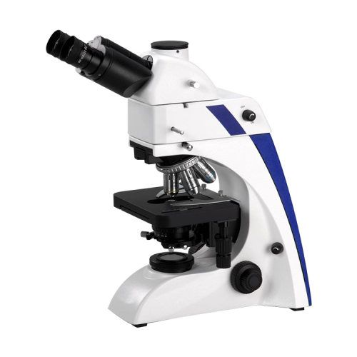
Bright-Field Microscope
How it works
Shines white light through the sample. You see the contrast between the sample and the background.
Pros
- Simple and affordable
- Great for stained slides
- Easy to operate
Cons
- Poor for transparent or live cells
- Low contrast without staining
Key Specs
- Magnification: 40x–1000x
- Best with stained or fixed samples
Best For
Teaching labs, tissue slides, and bacteria smears.
Dark-Field Microscope
How it works
Uses a special light stop so only scattered light from the specimen enters your eyes. The background looks black, sample looks bright.
Pros
- Highlights tiny, transparent things
- Great for live micro-organisms
Cons
- Hard to interpret for beginners
- Sensitive to dust and alignment
Key Specs
Very high contrast for small particles
Best For
Live bacteria, protozoa, and spirochetes.
Phase-Contrast Microscope
How it works
Converts differences in light phase into contrast perfect for transparent samples without staining.
Pros
- Perfect for live cells
- No staining required
- High contrast
Cons
- Halo artifacts around objects
- Costlier than bright-field
Key Specs
Designed for a transparent biological sample.
Best For:
Cell culture, live cells, organoids.
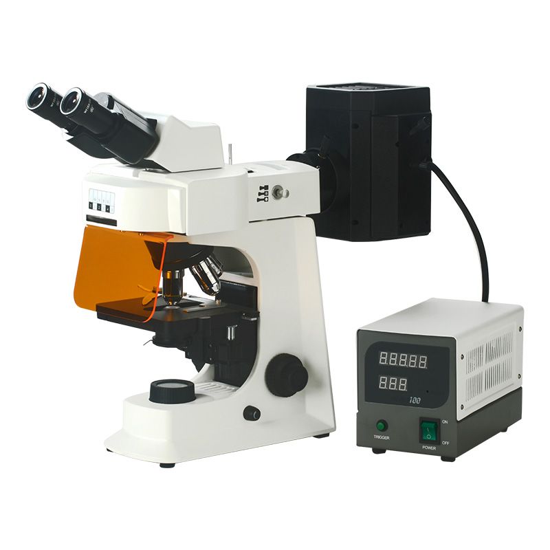
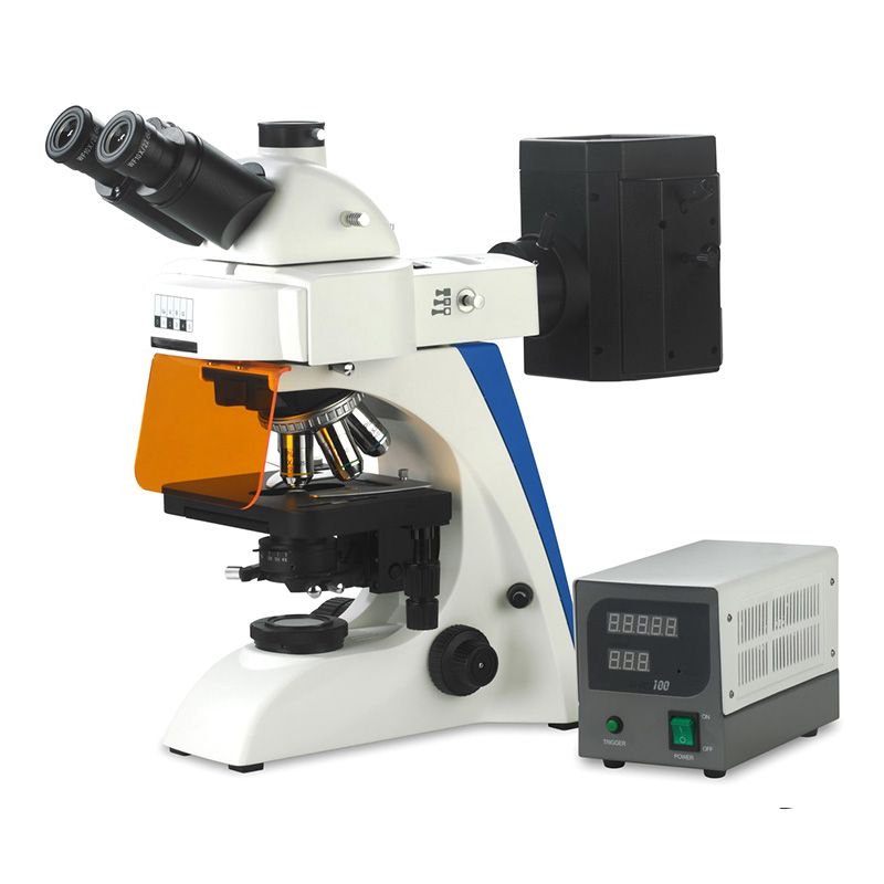
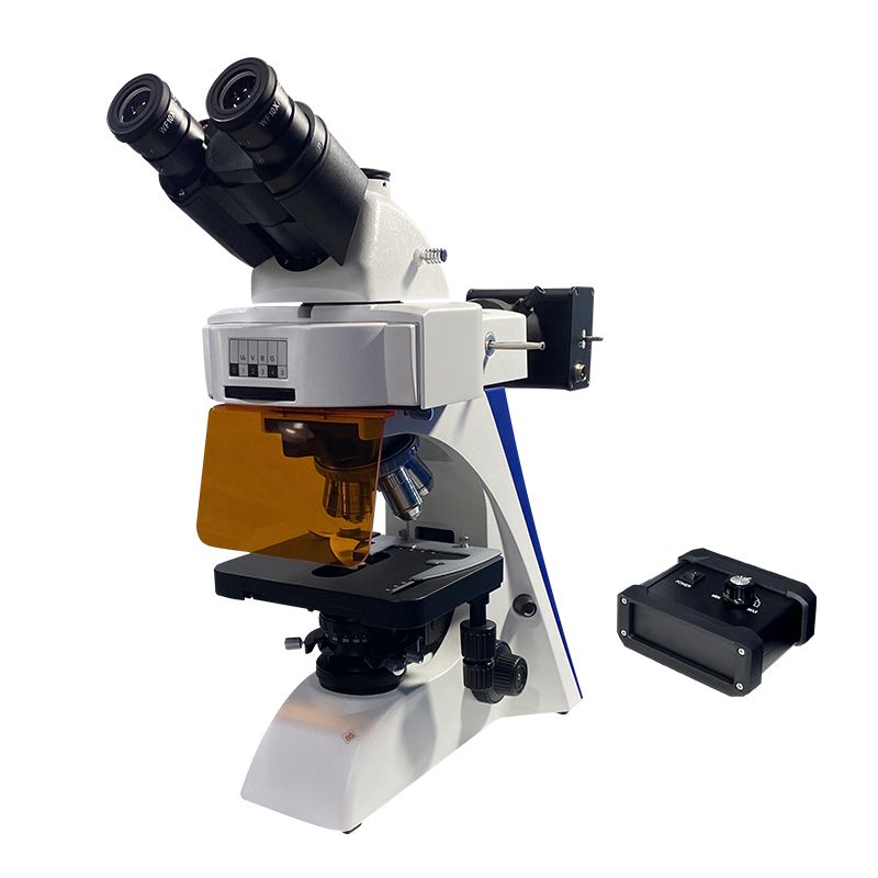
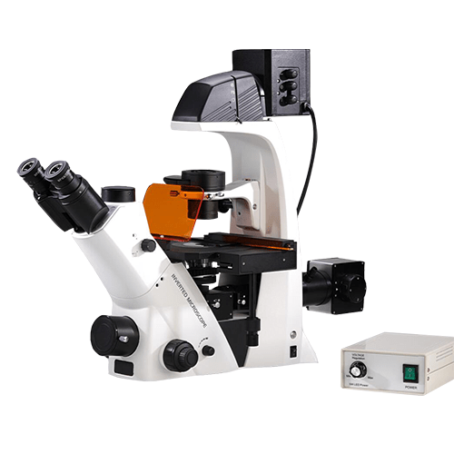
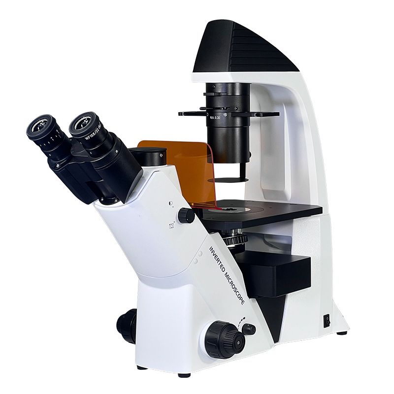
Fluorescence Microscope

How it works
Uses special light to excite fluorescent dyes or proteins. Only tagged structures light up.
Pros
- Super high contrast
- Can highlight specific proteins or organelles
- Works great for imaging cell functions
Cons
- Needs fluorophores (dyes)
- Bleaching and fading can occur
Key Specs
- Detects light emission rather than reflection
- Can image multiple colors
Best For
Immunofluorescence, gene expression, microbiology, diagnostics.
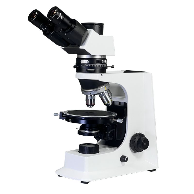
DIC / Nomarski Microscope
How it works
Uses polarized light and prisms to create a 3D-like shadow effect.
Pros
- Gorgeous, high-contrast images
- Works with live samples
- No staining
Cons
- Expensive
- Needs special optical components
Key Specs
- 3D-like visualization
- Great for transparent samples
Best For
Cell membranes, thin materials, microstructures.
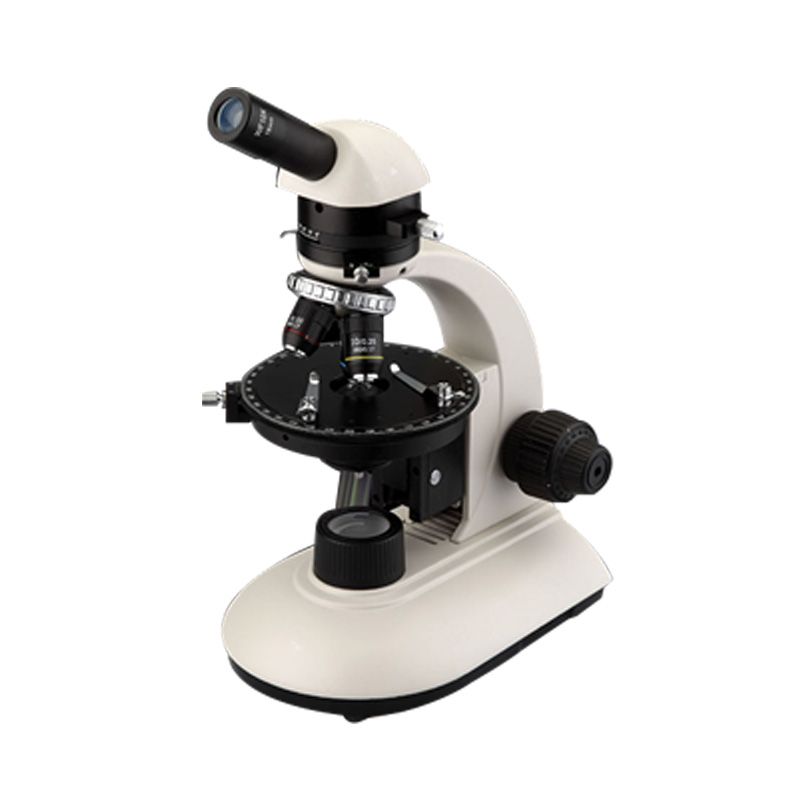
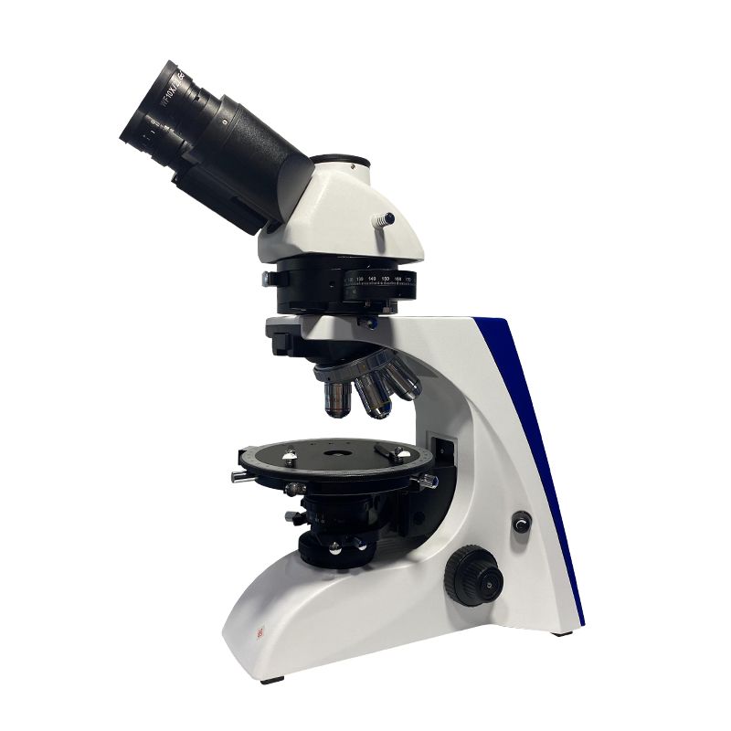
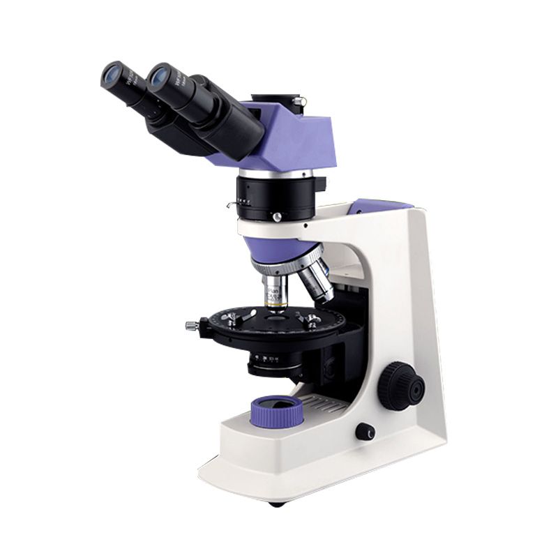
Polarizing Microscope

How it works
Uses polarized light to analyze structures that react to polarization.
Pros
- Reveals hidden crystal orientations
- Essential for geology and chemistry
Cons
- Not useful for most biological samples
Key Specs
- Detects birefringence
- Rotatable stage
Best For
Minerals, fibers, crystals, polymers.
Confocal Laser Scanning Microscope
How it works
Uses laser light + optical sectioning to create sharp, 3D images.
Pros
- High-resolution
- 3D imaging
- Eliminates out-of-focus blur
Cons
- Expensive
- Slower than basic light microscopes
- Needs fluorescent samples
Key Specs
- True optical sectioning
- 3D reconstruction
Best For
3D tissue samples, thick specimens, neuroscience, and cell biology.
How to Choose the Right Light Microscope
Here’s the quick, no-nonsense decision guide:
- For basic lab work or teaching: Bright-field
- For tiny, moving microorganisms: Dark-field
- For live, transparent cells: Phase-contrast or DIC
- For specific proteins or cell labeling: Fluorescence
- For crystals, minerals, fibers: Polarizing
- For 3D imaging or thick samples: Confocal
The easiest rule of thumb:
- If your sample is transparent: phase contrast / DIC
- If your sample glows: fluorescence / confocal
- If your sample is mineral or crystal: polarizing
- If you just need general observation: bright-field
Summary
Light microscopes come in different types because samples behave differently under light.
Each of the seven types solves a specific problem:
- Bright-field: everyday use
- Dark-field: tiny live organism
- Phase-contrast: transparent live cells
- Fluorescence: tagged molecules
- DIC : 3D-like transparent samples
- Polarizing: minerals & crystals
- Confocal: high-resolution 3D imaging
Choosing the right microscope simply depends on what you’re trying to see.