As life science research continues to advance, cell culture techniques have rapidly evolved from traditional 2D monolayer cultures to more complex 3D models (such as organoids and spheroids). These 3D models more realistically mimic the in vivo microenvironment and hold great promise for drug screening, disease modeling, and regenerative medicine. However, these advancements in culture technology also present new challenges for microscopic imaging: thicker samples, deeper imaging depths, and reduced phototoxicity are key challenges researchers must address.
An unsuitable microscope can lead to several major research pitfalls:
- You might miss subtle, yet crucial, structural changes deep within a 3D organoid.
- You’ll be unable to perform long-term live-cell imaging, losing dynamic data on cellular responses.
- You risk contamination or cell shock by manually removing cultures from a shaker incubator or CO₂ incubator for viewing.
At Scopelab, with years of experience in life science imaging, we understand that choosing the right microscope is crucial to achieving successful research. This article will systematically explain the differences in imaging requirements between 2D and 3D cell culture and provide a practical selection guide to help you make informed decisions in this rapidly evolving field.
2D vs. 3D Culture: The Imaging Requirement Divide
The first step in choosing your equipment is understanding the core imaging challenges presented by your culture model. Traditional 2D and advanced 3D systems place vastly different demands on your inverted microscope.
2D Culture: The Foundation
In 2D monolayer culture, cells are spread flat. Imaging is relatively simple, focusing on surface-level assessment. Your primary goal is to perform quick quality checks without high-level optical complexity.
3D Culture: Challenges of Depth and Dynamics
3D models (spheroids, organoids, hydrogels) are thick. This complexity creates two major hurdles: light cannot easily penetrate the sample, causing blur, and the cells require constant environmental control for long-term viability. This necessitates specialized equipment that allows for both depth and stability during imaging.
The table below summarizes the key differences in requirements and challenges:
| Feature | 2D Monolayer Culture (Standard) | 3D Culture (Advanced) |
| Primary Goal | Assess cell morphology, growth, and basic contamination. | Analyze complex structure, function, and dynamic processes. |
| Sample Thickness | Single cell layer (Thin). | Multiple cell layers; 50 μm to 500 μm+ (Thick). |
| Core Challenge | Routine quality control and manual handling risk. | Light scattering (blur) and maintaining viability. |
| Microscope Type | Inverted Phase Contrast Microscope. | Inverted Fluorescence Microscope with Z-stack capability or Confocal System. |
| Environmental Control | Not essential during the brief viewing period. | Mandatory: Requires a Stage-Top Incubator for stable live-cell imaging. |
| Key Equipment | Standard Inverted Microscope. | Motorized Inverted Microscope, Long Working Distance Objectives, and Environmental Chamber. |
| Required Data | Confluency, viability (basic). | Volume, cellular communication, dynamic drug response. |
If your research is moving toward 3D models, you must transition from a basic viewing device to an integrated live-cell imaging system that solves both the optical depth and environmental stability challenges.
Choosing Your Microscope: Technology Tiers
Your budget and research goals will determine which technological tier you need to invest in, and Scopelab has solutions for every stage of your cell culture research.
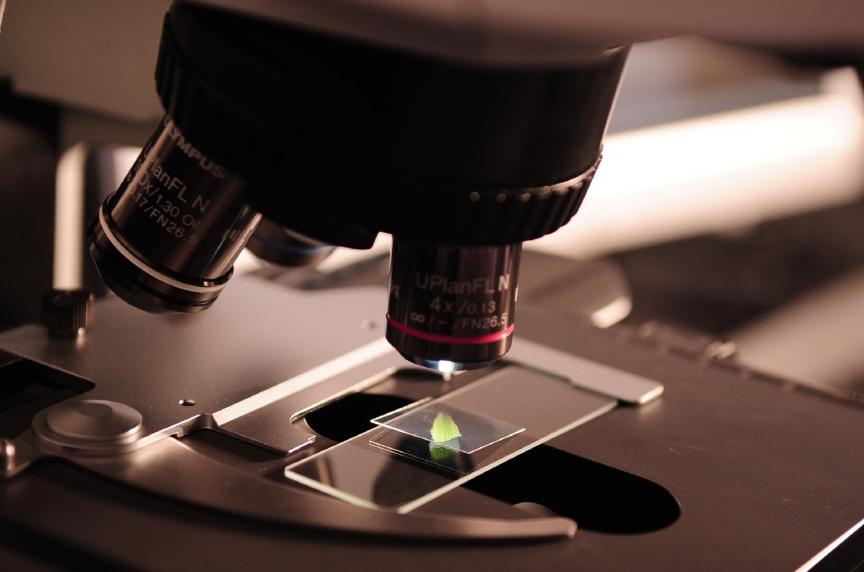
Tier 1: Routine Assessment – The Inverted Phase Contrast Microscope
This is the essential starting point for every lab. It uses an optical technique (phase contrast) to create high-contrast images of transparent, unstained cells. This instrument is the daily workhorse for all cell culture lab equipment.
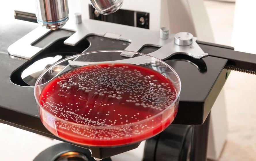
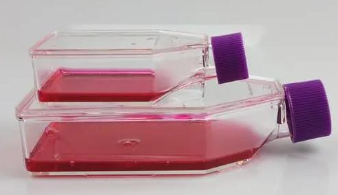
Best For: Everyday checks of 2D cell morphology, confluence, and quick checks of contamination in standard flasks and plates.
Limitation: Provides little insight into the internal structure of thick 3D cultures.
Scopelab’s Recommendation: Our standard Inverted Phase Contrast Microscope offers excellent optical quality and a spacious stage to easily accommodate large multi-well plates and T-flasks—perfect for daily 2D work. We prioritize a robust build and high-brightness LED illumination for clear viewing.
Tier 2: Molecular Analysis – The Inverted Fluorescence Microscope
Adding fluorescence capability allows you to target and visualize specific molecules, proteins, or cellular events using fluorescent dyes (e.g., assessing viability with a live/dead stain). This upgrade is necessary for detailed analysis beyond simple morphology.
Best For: Evaluating apoptosis, assessing transfection efficiency, and visualizing fluorescent markers in the surface layers of 3D structures.
Upgrade Highlight: Requires high-quality LED light sources and high-transmission filter cubes to capture weak signals.
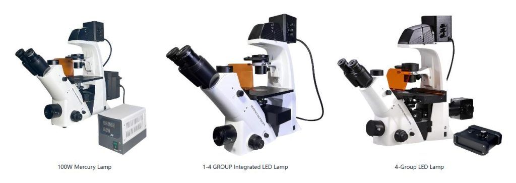
Scopelab’s Recommendation: Upgrade your system with our modular LED Fluorescence Illumination System. This can be added to your base inverted microscope and provides multiple LED wavelengths and a motorized filter turret. This flexibility allows you to perform multi-channel imaging on your 3D cell culture samples. Or directly choose our BDS500-FL Inverted Fluorescence Microscope can meet your needs.

Tier 3: The Gold Standard – Live-Cell Imaging Integrated Systems
To overcome the inherent limitations of 3D imaging and maintain cell viability, you must combine a high-quality inverted microscope with a precise environmental control unit. This is where the term “inverted microscope for cell culture” takes on its most advanced meaning—it is an integrated system designed for high-content analysis.
This system is engineered for reliable, high-resolution dynamic data acquisition:
| Component | Function in 3D Culture | CQ Scope Lab Advantage |
| Stage-Top Incubator | Provides stable temperature, CO₂ and humidity control directly on the microscope stage, enabling long-term live-cell imaging. | Our Stage-Top Live Cell Incubator ensures precise control (±0.1 ℃), mimicking your dedicated CO₂ incubator environment for experiments lasting days. |
| Motorized Z-Drive | Allows optical sectioning (acquiring image stacks) to reconstruct a crisp, 3D view of the organoid, eliminating out-of-focus blur. | Seamless Z-axis control for precise multi-layer image acquisition and 3D reconstruction. |
| Long Working Distance Objectives | Essential for focusing through thick plastic plates, hydrogels, and 3D scaffolds. | We supply a range of specialized objectives critical for successful 3D cell culture viewing. |
Scopelab’s Recommendation: Our Live-Cell Imaging Integrated System (similar to the specialized setups featured on CQ Scope Lab’s application pages) is the complete solution. It integrates all the necessary environmental, optical, and mechanical components. Whether tracking cell migration or monitoring the effect of a drug on a 3D cell culture over 48 hours, this system provides the stability and resolution needed for translational research.
Final Considerations: Choosing Your Cell Culture Equipment Partner
Acquiring an advanced microscope is an investment in your research’s future. When selecting your provider, ensure they meet these criteria:
- Application Expertise: Does the supplier understand the complexities of 3D animal cell culture and the specific needs of inverted microscope operation with environmental control?
- Scalability: Can the system grow with your lab, easily integrating modules like a CO₂ controller or advanced image analysis software?
- Support and Training: High-end systems require professional installation and training to maximize your investment.
At Scopelab, we don’t just sell cell culture lab equipment; we provide end-to-end solutions. Our technical team specializes in both basic inverted microscopes and complex live-cell imaging setups, ensuring your transition to advanced 3D research is seamless and successful.
Contact Scopelab today to schedule a consultation and see how our tailored imaging solutions can elevate your cell culture research.