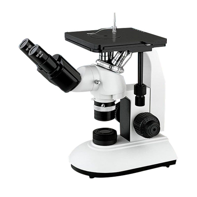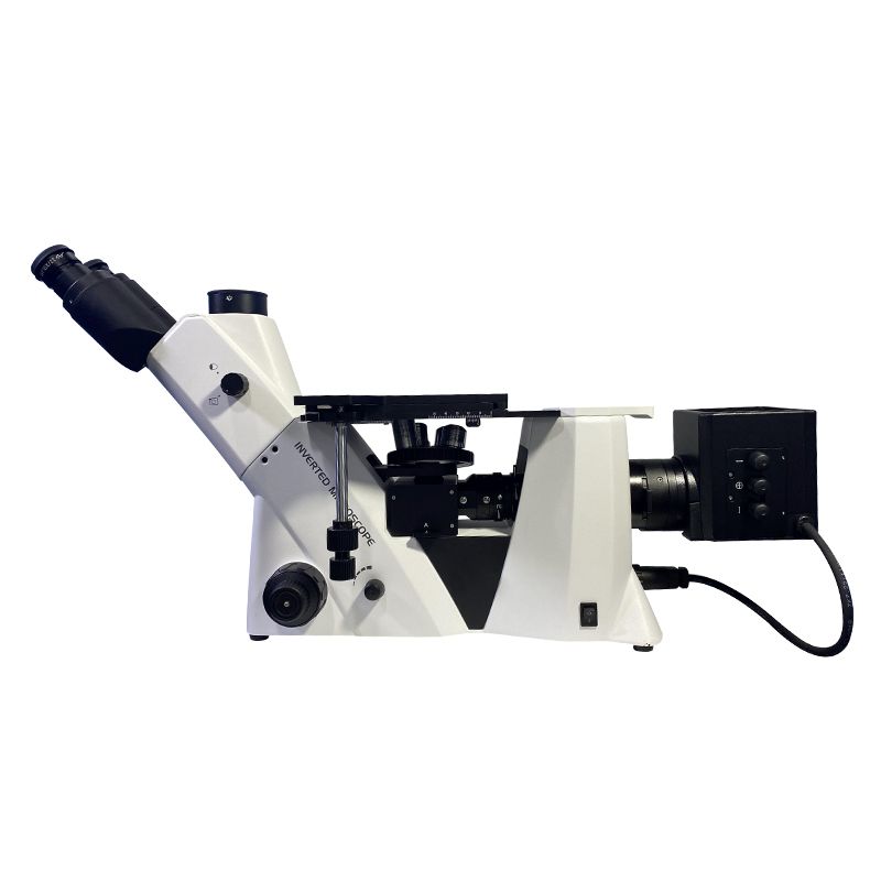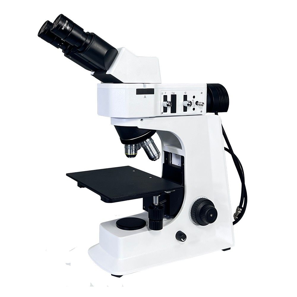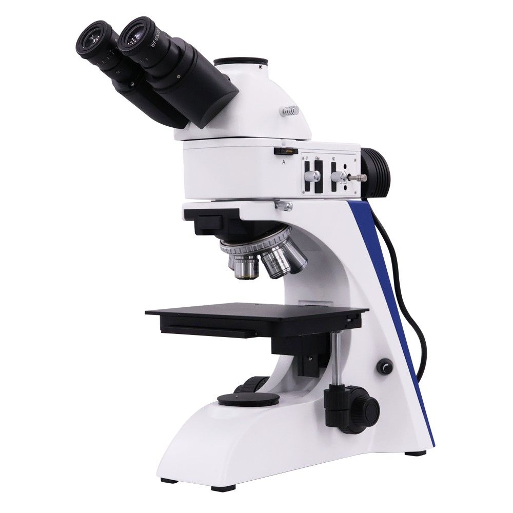
Metallurgical microscopes, also referred to as metallographic microscopes, typically use one of four common methods of observation to examine the physical characteristics of Metallurgical samples: bright field, dark field, polarized light, or differential interference. The latest laser confocal method appeared in the last ten years. With a bright field serving as the basic core, the other four functions can be thought of as “building blocks” that are assembled onto a metallurgical microscope, by the design concept of “modular design, building block structure” in modern instruments.
Method 1: Bright Field
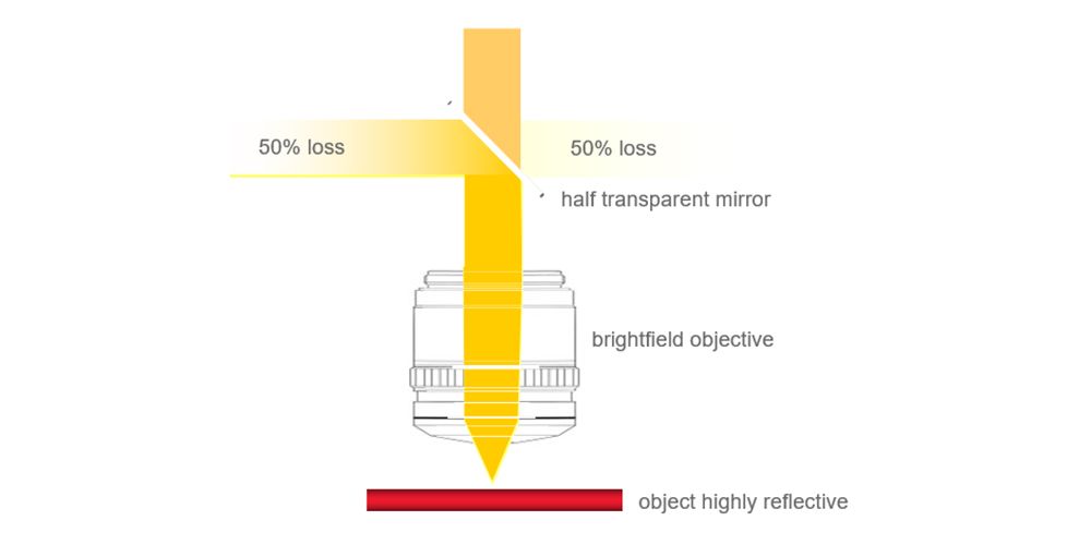
The primary method of observation used in Metallurgical research is bright field illumination because it is easy to use, adaptable, and offers good contrast for a wide range of samples. The sample surface is irradiated vertically or nearly vertically by the incident light, and the reflected light from the surface enters the objective lens for being imaged. “Bright field” illumination refers to the situation where a bright piece, such as a mirror sample, is in the field of view and the tissue on the sample reflects the colored image in the bright field of view. It is easy to use and doesn’t require any special equipment to use this illumination method. Additionally, it is versatile and can be used to examine a broad range of samples, such as polymers, metals, and ceramics.
Advantages of Bright Field
- High brightness and uniform field of view. High brightness is crucial as it permits greater light to enter the eyepiece, thereby producing a brighter image. In bright field illumination, where samples are frequently opaque or have low contrast, this is especially crucial. It is simpler to see small details and defects in the sample with a brighter image.
- Wide range of applications. A vast variety of samples, such as metals, ceramics, polymers, and biological specimens, can be observed using it. This is because bright field illumination doesn’t call for any particular sample handling or preparation.
- Simple operation and low price. It doesn’t require adjustments or specialized equipment. The objective lens creates the image, while the light source is only focused on the sample.
Disadvantages of Bright Field
- low-contrast specimens have low contrast. To produce contrast, bright field illumination uses variations in the way that light is absorbed or scattered across various sample features. A small difference in light absorption or scattering will result in a small contrast in the image.
- Specimens have no three-dimensional effect. The objective lens creates the image in bright field illumination, which uses a single light source to illuminate the sample. As a result, even though the sample is three-dimensional, a two-dimensional (2D) image of it is produced.
Method 2: Dark Field
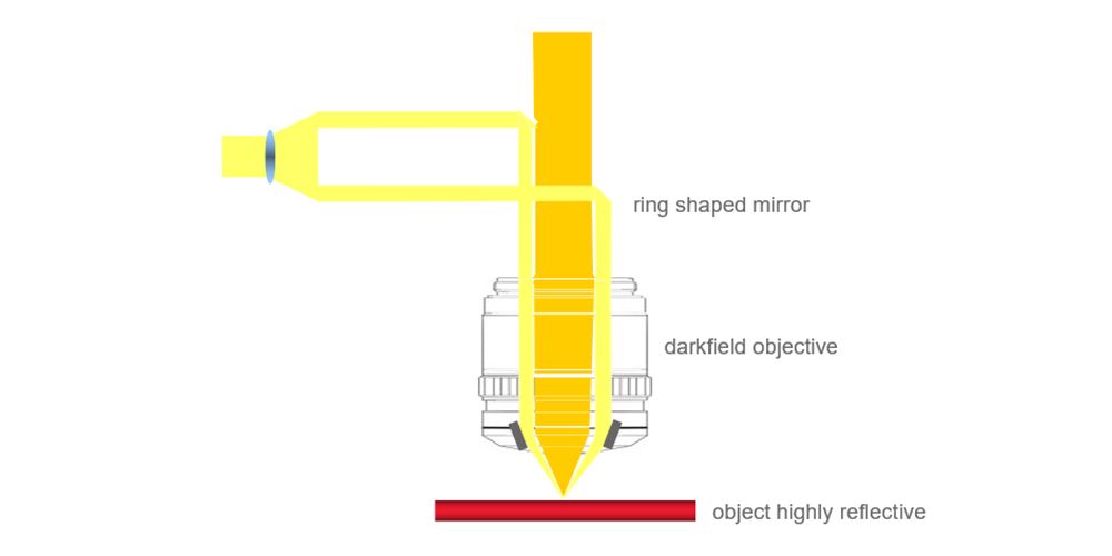
The objective lens’s periphery illuminates the sample, preventing illumination light from entering the lens itself. This allows for the formation of an image on the sample’s surface due to diffuse reflection light. If the sample has a mirror surface, light reflected from it will still be reflected in the opposite direction at a steep angle, making it impossible for light to enter the objective lens and leaving the field of view completely dark. “Dark field” illumination refers to the process whereby the objective lens reflects the tissue on the sample with a bright white image in the dark field of view, like the stars in the night sky.
Advantages of Dark Field
- Observe extremely small objects with a resolution of 0.02-0.004um (0.4um in bright field). Smaller objects can be observed with dark field illumination because it has a higher resolution than bright field illumination. This is because dark field illumination only displays light that the sample has scattered. Even from very small objects, this scattered light is frequently much brighter than the direct light that flows through the sample.
Disadvantages of Dark Field
- Only the existence, movement, and external form of objects can be observed. This is because light scattered by the sample is the only light visible under dark field illumination. The internal structure of the sample cannot be inferred from this dispersed light.
Method 3: Polarizing
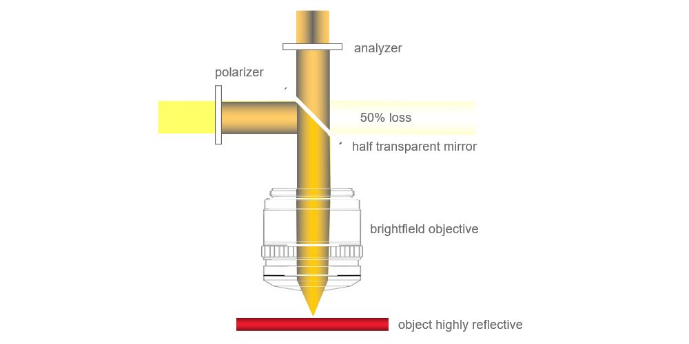
Electric light and sunlight are considered forms of natural light. Its light vibration is perpendicular to the direction of propagation and balanced in all directions. Polarized light is defined as light whose vibration is restricted to a single direction in the plane perpendicular to the direction of propagation. Two common methods for obtaining polarized light are using an artificial polarizer or a polarizing prism.
Advantages of Polarizing Light Microscope
- Improved contrast: Using a polarized light microscope, birefringent sample contrast can be greatly enhanced. This is because polarized light will be split into two beams with distinct polarizations by birefringent samples. The sample can then be seen in high contrast by filtering these two beams.
- Ability to reveal defects: By using a polarized light microscope, flaws in samples that are invisible in bright field illumination can be found. Flaws may exhibit birefringence that differs from that of the surrounding material.
- Ability to identify crystal structures: The crystal structures of minerals and other materials can be identified using a polarized light microscope. A material’s crystal structure determines its birefringence.
Disadvantages of Polarizing Light Microscope
- More complex setup: A more complex setup is needed for a polarized light microscope than for bright field illumination. You need both an analyzer and a polarizer.
- Less versatile: Bright field illumination offers greater versatility than the polarized light microscope. The fact that only birefringent samples can be observed using a polarized light microscope.
- Difficulties in interpreting images: Particularly for novice users, polarized light microscope images can be challenging to interpret.
Method 4: Differential Interference (DIC)
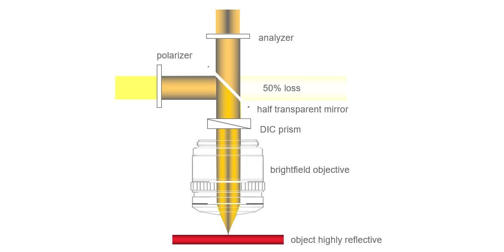
Polarized light interference is the basis for differential interference. The illumination light transforms into two beams of diffracted light from a differential interference contrast prism, and the sample height difference results in a tiny difference in the optical path that eventually transforms into a polarizer and a differential interference contrast prism. dark and light contrast.
Advantages of Differential Interference
- It can give the item being examined a three-dimensional sense.
- The observation effect makes greater sense.
- An objective lens of specialization is not required, and fluorescence observation yields better results.
- To attain the desired effect, one can modify the color changes of the object and background.
Disadvantages of Differential Interference
- Birefringent materials cannot produce DIC microscope effects and cannot be used to observe cultures in plastic containers. High light intensity is necessary. The adjustment for directional microscope sensitivity is complicated.
Conclusion
Knowing how to use Metallurgical microscopes for observation is essential since it gives us access to a variety of data regarding the microstructure of metals. Every observation method has advantages and disadvantages, so it’s critical to select the best approach for the given situation. The basic observation technique is bright field illumination, which is used to examine a variety of supplies. A more specialized observation technique called dark field illumination is used to enhance the contrast and depth of field in pictures. Studying birefringent samples requires the use of a specialized observation technique called polarized light illumination. A specialized observation technique called differential interference contrast (DIC) microscope is used to create three-dimensional (3D) images of samples. We can select the best method for a given application and get the information we require by being aware of the various Metallurgical microscope observation techniques.