A stereo microscope (also known as a stereo microscope or dissecting microscope) is a light microscope designed for low-magnification observation. It doesn’t penetrate deeply into the microscopic world, so you can’t see cells. Instead, it provides a 3D view of objects that are almost as visible to the naked eye, but with greater detail. It’s an excellent tool for studying surfaces, structures, and morphology. If your primary goal is to study cells, you’ll need a compound microscope, not a stereo microscope.
What exactly is a stereo microscope?
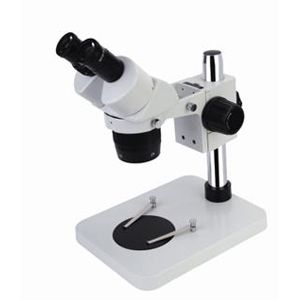
A stereo microscope is a low-magnification optical microscope that uses conventional visible light. Unlike the high-magnification biological microscopes we often associate with stereo microscopes, it is not designed to view cells or bacteria. Instead, it is used to observe three-dimensional details on the surface of an object at relatively low magnification (usually 10–80x). Therefore, a stereo microscope is not used to “see cells in the microscopic world” but to “see objects that are barely visible to the naked eye and magnify them to a more three-dimensional and clearer level.”

The biggest features of a stereo microscope
- 3D imaging: Because each eye has a separate optical path, it’s like wearing 3D glasses.
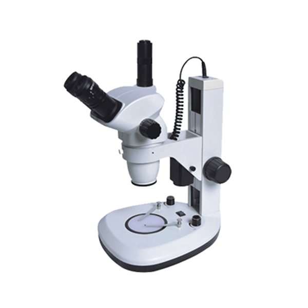
- Large working distance :There’s ample space between the lens and the sample, allowing for direct manipulation of the sample (e.g., dissection, welding, repair).
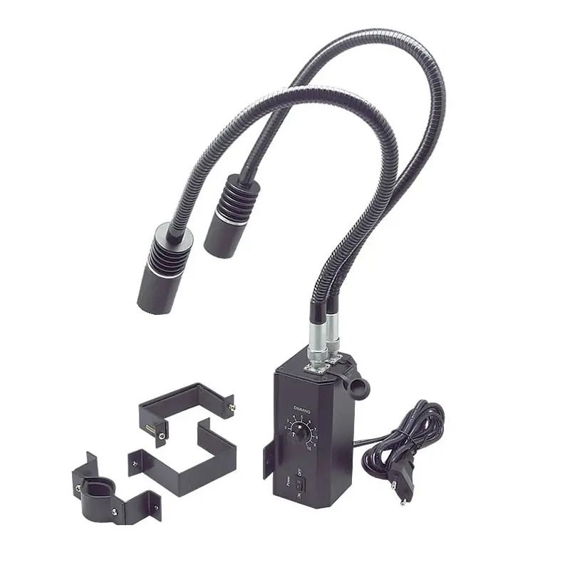
- Flexible light source: Can use either reflected light (illuminating the surface) or transmitted light (illuminating through thin samples).
Why “stereo”? Because it uses two separate optical paths, one for each eye. This creates a three-dimensional view—almost like wearing 3D glasses. Therefore, stereo microscopes are also 3D.
- Reflected light (incident light): Illuminates opaque objects such as insects, rocks, and circuit boards.
- Transmitted light: Passes through thin or translucent samples, such as thin plant parts or small aquatic organisms.
What is a stereo microscope best for viewing?
“Stereo microscopes are best suited for observing large, opaque specimens where surface detail and a sense of space are important.” Stereo microscopes are used to “see details clearly and give a sense of three-dimensionality.” This is the appeal of stereo microscopes: they are excellent for observing surface details and 3D structures. Think of them as a super-powerful magnifying glass.
- Stereo microscope magnification: 10x–80x (sometimes up to 100x).
- However, the magnification required to clearly observe cells is 400x–1000x (compound microscope range).
If your primary goal is to study cells, you will need a compound microscope, not a stereo microscope.
Some common uses include:
- Biology: Insects, plant parts, small animals, dissection
- Electronics: Soldering, PCB inspection, microchips
- Jewelry and gemstones: Clarity inspection, setting gemstones
- Geology: Rocks, minerals, crystals
- Everyday objects: Coins, stamps, tiny mechanical parts
Stereo Microscope vs. Compound Microscope
| Feature | Stereo Microscope | Compound Microscope |
| Magnification | 10x–80x (low) | 40x–1000x+ (high) |
| View | 3D | 2D |
| Light Source | Reflected + Transmitted | Mainly Transmitted |
| Best For | Surface detail, solid 3D objects | Cells, tissues, bacteria |
| Samples | Insects, plants, rocks, PCBs, jewelry | Thin slices, transparent samples |
| Can See Cells? | Not clearly | Yes |
How to Choose the Right Microscope
| Microscope Type | Main Features | Limitations | Best For / Research Objects |
| Stereo Microscope (Dissecting Microscope) | – Low magnification (10x–80x)- 3D view – Uses both reflected & transmitted light – Large working distance (can operate on samples while viewing) | – Cannot see cells or bacteria – Limited magnification | – Solid & opaque objects – Insects, plants, rocks, jewelry, PCBs, coins |
| Compound Microscope (Biological Microscope) | – High magnification (40x–1000x)- 2D image – Uses transmitted light – Great for thin, transparent samples | – Cannot view large/thick samples – 2D only, no depth perception | – Cells, tissues, bacteria, microorganisms – Blood smears, onion skin, protozoa |
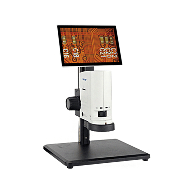
| Digital Microscope | – Built-in camera & screen – Easy sharing & image capture – Can be stereo – or compound-style | – Image quality depends on sensor – May lack true optical depth | – Education, demonstrations, quality control – General hobby use |
| Electron Microscope (SEM/TEM) | – Extremely high magnification (up to millions of times) – Can see ultrastructure, organelles, nanoscale features – SEM = 3D surface view – TEM = internal structures (2D slices) | – Very expensive – Requires special sample prep – No live specimens (samples often coated or fixed) | – Viruses, nanomaterials, organelles – Advanced research in biology & materials |
| Polarizing Microscope | – Uses polarized light – Highlights crystal orientation & optical properties | – Limited to anisotropic materials – Not useful for cells | – Minerals, crystals, geology, forensic samples |
| Fluorescence Microscope | – Uses special light & dyes – Allows visualization of tagged structures – Can highlight specific proteins or organelles | – Expensive – Requires staining/fluorescent dyes | – Cell biology, microbiology – Medical & molecular research |
Summary
Finally, can a stereo microscope see cells? No. Stereo microscopes are designed for low magnification (10x to 80x) and can provide a 3D view of solid objects—ideal for studying insects, plants, rocks, jewelry, or circuit boards, but they aren’t powerful enough to visualize cells or bacteria.
If your goal is to explore the microscopic world of cells and microorganisms, you’ll need a compound microscope (40x to 1000x, 2D view, transmitted light). For more advanced research, tools like fluorescence or electron microscopes can open up even finer perspectives.
- Stereo microscopes are best for observing 3D surface details of larger, opaque objects.
- Compound microscopes are best for observing thin, transparent specimens like cells and tissues.
- Other microscopes are specialized for materials science, nanotechnology, or advanced biology.