From examining cellular structures to exploring nano-level phenomena, microscopes have become essential tools across various scientific disciplines, play an indispensable role in unraveling the mysteries of the microscopic world. With advancements in technology, digital cameras have revolutionized the way we capture and analyze microscopic images. In this article, we delve into the world of digital cameras for microscope analysis, exploring their types and factors to consider when selecting the right one for your research needs.
Understanding Microscope Imaging
What are Microscopes and How does Microscope Imaging Work?
Microscopes are powerful instruments designed to magnify and visualize objects that are too small to be seen by the naked eye. They function based on the principles of optics, utilizing lenses to bend and focus light rays. There are several types of microscopes, including optical microscopes, electron microscopes, and scanning probe microscopes, each with its own unique capabilities and applications.
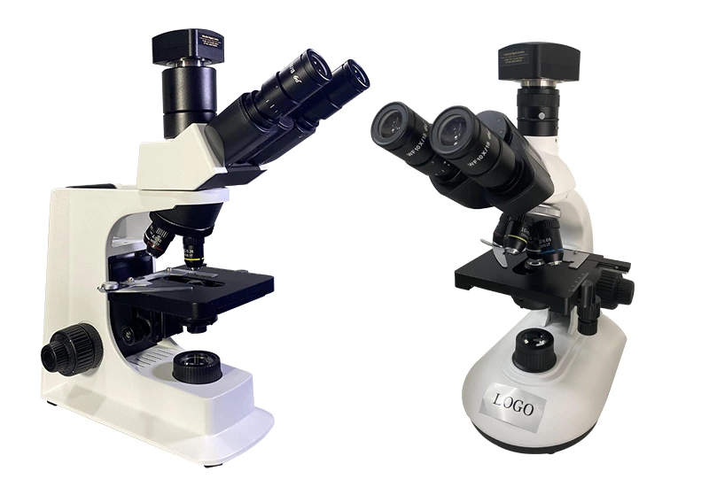
Microscope imaging involves capturing high-resolution images of tiny specimens magnified under a microscope. Traditionally, this process relied on eyepieces and photographic film to document observations. However, digital cameras have transformed microscopy by enabling real-time imaging, precise documentation, and digital storage of images. These cameras are seamlessly integrated with microscopes, providing researchers with powerful tools for analysis and documentation.
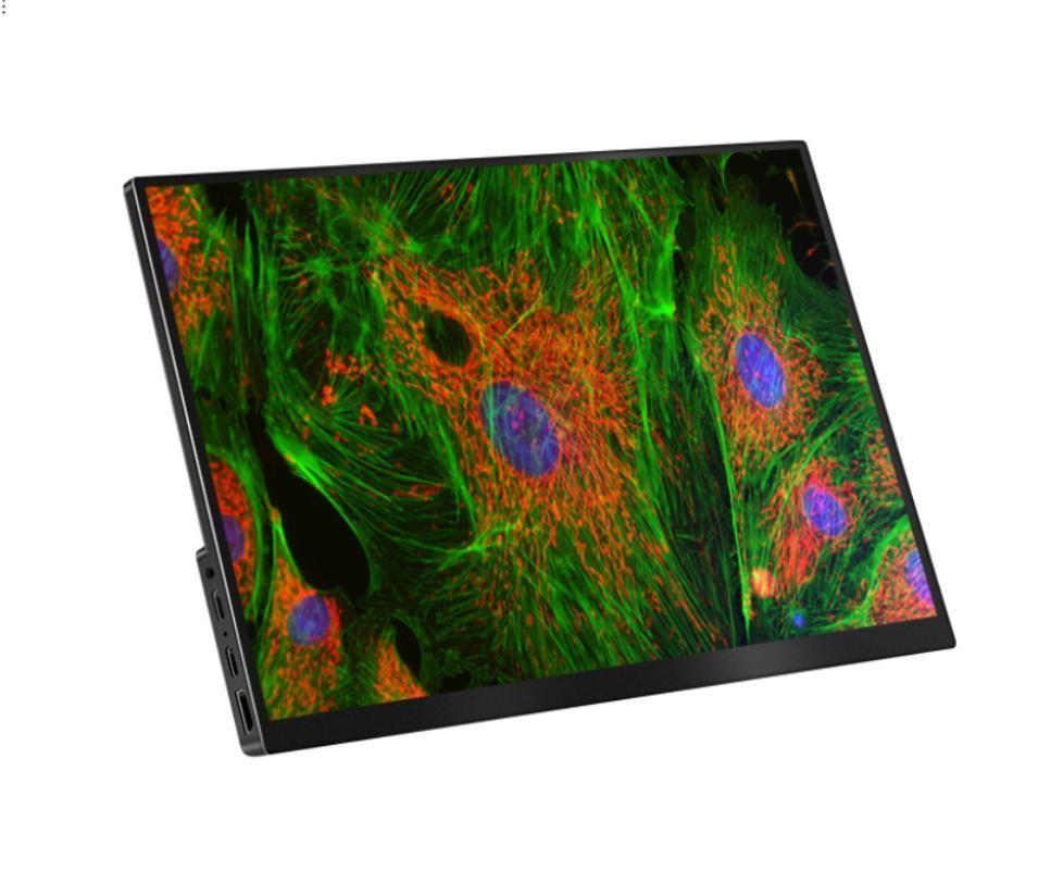
High-quality imaging is paramount in microscopy as it directly impacts the accuracy and reliability of observations and analyses. Clear, sharp images are essential for identifying cellular structures, discerning fine details, and making precise measurements. In research and clinical settings, the ability to capture high-quality images is critical for studying biological samples, diagnosing diseases, and advancing scientific knowledge.
Why Choose Digital Cameras?
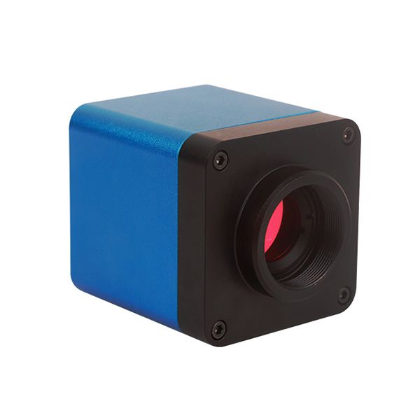
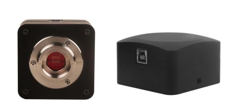
Digital cameras play a pivotal role in capturing and enhancing microscope images, replacing traditional film-based photography with advanced digital imaging technology. These cameras are equipped with sensors that convert light into electronic signals, which are then processed and converted into digital images. By integrating digital cameras with microscopes, researchers can capture real-time images, record videos, and perform quantitative analysis with unprecedented precision and efficiency.
Moreover, digital cameras offer several advantages over traditional methods of microscopy imaging:
- Instant Image Acquisition: Unlike traditional film-based photography, digital cameras allow for instant image acquisition. Researchers can view and capture images in real-time, enabling immediate analysis and adjustments.
- Electronic Storage and Sharing: Digital images can be stored electronically on computer systems, external drives, or cloud storage platforms. This eliminates the need for physical storage space and simplifies data management. Additionally, digital images can be easily shared with collaborators or published online, facilitating collaboration and dissemination of research findings.
- Enhanced Image Quality: Digital cameras can produce high-resolution images with exceptional clarity and detail. Advanced sensor technology and image processing algorithms enhance image quality by reducing noise, improving contrast, and optimizing color accuracy.
- Quantitative Analysis: Digital images can be analyzed quantitatively using specialized software tools. Researchers can measure dimensions, quantify intensities, and perform statistical analysis directly on digital images, enabling precise and objective data analysis.
- Image Manipulation and Enhancement: Digital images can be manipulated and enhanced using image processing software. Researchers can adjust brightness, contrast, and color balance, as well as apply filters and overlays to enhance specific features or highlight areas of interest.
- Real-Time Imaging: Digital cameras enable real-time imaging, allowing researchers to capture dynamic processes and events as they occur. This is particularly useful for studying live cells, fast-moving specimens, and time-sensitive experiments.
- Cost-Effectiveness: While the initial investment in digital cameras may be higher compared to traditional film-based systems, digital imaging offers long-term cost savings. Digital cameras eliminate the recurring costs associated with film processing and development, as well as the need for consumable materials such as film rolls and chemicals.
- Remote Access and Collaboration: Digital imaging technology enables remote access to microscope images, allowing researchers to view and analyze samples from anywhere with an internet connection. This facilitates collaboration between researchers located in different geographical locations and enables remote supervision and training.
Overall, digital cameras offer significant advantages over traditional methods of microscopy imaging, including instant image acquisition, electronic storage and sharing, enhanced image quality, quantitative analysis capabilities, real-time imaging, cost-effectiveness, and remote access and collaboration. These advantages have revolutionized microscopy imaging, enabling researchers to conduct more efficient, precise, and impactful scientific research.
Types of Digital Cameras for Microscope Analysis
Digital cameras for microscope analysis come in various types, each offering distinct features and capabilities tailored to different research needs. Here are the main types:
CCD (Charge-Coupled Device) Cameras:
CCD cameras have been a staple in microscopy imaging for many years.
- They utilize a grid of light-sensitive pixels to capture images, converting light into electronic signals.
- CCD cameras are known for their high sensitivity and low noise, making them well-suited for low-light microscopy applications such as fluorescence microscopy.
- They offer excellent image quality with accurate color reproduction and low levels of image distortion.
- CCD cameras are ideal for capturing static images and are commonly used in research laboratories and clinical settings.
CMOS (Complementary Metal-Oxide-Semiconductor) Cameras:
CMOS cameras have gained popularity in recent years due to advancements in technology.
- They operate similarly to CCD cameras but use a different type of sensor technology.
- CMOS cameras offer advantages such as lower cost, higher frame rates, and lower power consumption compared to CCD cameras.
- They excel in dynamic imaging applications, including live cell imaging, time-lapse microscopy, and high-speed imaging.
- CMOS cameras provide comparable image quality to CCD cameras, with improvements in sensor technology narrowing the performance gap between the two.
sCMOS (Scientific Complementary Metal-Oxide-Semiconductor) Cameras:
sCMOS cameras represent the latest advancement in digital camera technology for microscopy.
- They combine the best features of CCD and CMOS cameras, offering high sensitivity, low noise, and fast frame rates.
- sCMOS cameras are particularly well-suited for demanding microscopy techniques such as super-resolution microscopy, single-molecule imaging, and high-speed imaging.
- They provide researchers with superior image quality, enhanced sensitivity, and improved dynamic range, enabling groundbreaking discoveries in various fields of science.
- While sCMOS cameras tend to be more expensive than CCD and CMOS cameras, their exceptional performance and versatility justify the investment for many research applications.
These are the primary types of digital cameras used for microscope analysis, each offering unique features and advantages. Here’s a table outlining the features, pros, cons, and applications of the three main types of digital cameras for microscope analysis:
| Type | Features | Pros | Cons | Applications |
| CCD Cameras | Utilizes charge-coupled device (CCD) sensor technology | High sensitivity | Lower frame rates compared to CMOS and sCMOS cameras | Fluorescence microscopy Low-light microscopy Static imaging |
| Excellent color reproduction | Low noise | Higher cost compared to CMOS cameras | Histology Pathology Clinical diagnostics | |
| Good dynamic range | ||||
| CMOS Cameras | Utilizes complementary metal-oxide-semiconductor (CMOS) sensor technology | Lower cost compared to CCD cameras | Lower sensitivity compared to CCD and sCMOS cameras | Live cell imaging Time-lapse microscopy High-speed imaging |
| Higher frame rates | Lower power consumption | Limited dynamic range compared to sCMOS cameras | Cell biology Neuroscience Biophysics | |
| Lower noise | Less heat generation | Relatively recent technology, may have fewer options available | ||
| sCMOS Cameras | Utilizes scientific complementary metal-oxide-semiconductor (sCMOS) sensor technology | High sensitivity | Higher cost compared to CCD and CMOS cameras | Super-resolution microscopy Single-molecule imaging High-speed imaging |
| Low noise | Fast frame rates | Larger file sizes compared to CCD and CMOS cameras | Structural biology Nanotechnology Materials science | |
| Wide dynamic range | Improved image quality | May require powerful computer hardware for data processing |
These are general features, pros, cons, and applications associated with each type of digital camera for microscope analysis. Specific models may vary in their performance characteristics and suitability for particular applications. Researchers should carefully consider their imaging requirements and budget constraints when selecting a digital camera for their microscopy needs.
Factors to Consider When Choosing a Digital Camera
When selecting a digital camera for microscope analysis, several factors should be taken into consideration to ensure it meets the specific imaging requirements of your research or application. Here are some key factors to consider:
- Resolution: The resolution of the camera determines the level of detail captured in an image. Higher resolution cameras can resolve finer details and are essential for applications requiring precise measurements or detailed analysis.
- Sensitivity: Sensitivity refers to the camera’s ability to capture faint signals or low-light conditions. Cameras with high sensitivity perform well in low-light microscopy techniques such as fluorescence microscopy. Consider the sensitivity of the camera’s sensor, as well as factors like quantum efficiency and readout noise.
- Frame Rate: The frame rate of the camera dictates how quickly it can capture images. Higher frame rates are essential for dynamic imaging applications, such as live cell imaging or high-speed microscopy. Ensure the camera can capture images at a frame rate suitable for your specific application.
- Dynamic Range: Dynamic range refers to the range of light intensities that the camera can accurately capture. A wide dynamic range is important for preserving detail in both bright and dark areas of an image, especially in samples with high levels of contrast.
- Compatibility: Ensure that the camera is compatible with your microscope setup, including the camera port, mounting system, and software interface. Compatibility with microscopy software is crucial for seamless image acquisition, analysis, and documentation.
- Software and Image Processing: Consider the availability and capabilities of software for image acquisition, processing, and analysis. Look for features such as real-time image processing, measurement tools, and compatibility with your existing image analysis workflows.
- Cost: Digital cameras for microscopy vary widely in price, depending on their features, performance, and brand. Consider your budget constraints and balance them against the required features and performance characteristics of the camera.
- Brand and Support: Choose a reputable brand known for quality and reliability. Consider factors such as warranty, technical support, and availability of accessories or upgrades. Opting for a well-established brand can provide peace of mind and ensure ongoing support for your investment.
- Future Expandability: Anticipate future needs and consider whether the camera allows for upgrades or additional features. Some cameras may offer modular designs or compatibility with accessories such as motorized stages or filters, enabling future expansion of imaging capabilities.
- User Experience: Finally, consider the user experience and ease of use of the camera. Look for features such as intuitive controls, ergonomic design, and user-friendly software interfaces to streamline your imaging workflow and minimize training time for users.
By carefully considering these factors and evaluating your specific imaging requirements, you can choose a digital camera that meets your needs and enables high-quality microscopy imaging for your research or application.
Conclusion
Digital cameras have become indispensable tools for microscope analysis, enabling researchers to capture, analyze, and document microscopic images with unprecedented precision and efficiency. Whether you’re conducting fundamental research or clinical diagnostics, selecting the right digital camera is essential for achieving accurate and reliable results. By understanding the different types of cameras available and considering key factors such as resolution, sensitivity, and frame rate, researchers can make informed decisions that meet their specific imaging needs. If you are not sure to choose the proper type for your project, please feel free to contact Scopelab – the microscope manufacturer and supplier. They can provide you professional advices and microscope cameras.