

The biliary system is inherently complex due to the branched network structure of the bile ducts both inside and outside the liver. Bile can either enter the gallbladder for storage or be discharged directly into the duodenum via the common bile duct to participate in digestion.
A high-precision liver anatomy model is essential for helping students and clinicians understand how the gallbladder connects to the biliary tree. By using distinct color-coding, this model displays the complete vascular network of the liver—including the portal vein, intrahepatic bile ducts, and extrahepatic bile ducts—making bile drainage and critical surgical landmarks (such as the cystic duct and common bile duct) much clearer to visualize.
What is the Biliary Tree?
The biliary tree is a branching system composed of intrahepatic and extrahepatic bile ducts, with the primary function of transporting bile. It includes critical structures such as the left hepatic duct, right hepatic duct, common hepatic duct, cystic duct, and common bile duct. Together, these structures complete the collection, storage, and discharge of bile.
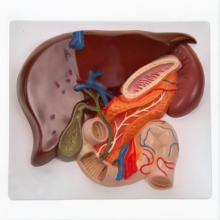
Biliary System Visualization: Hepatic and Cystic Ducts
The number one complaint educators have regarding low-end anatomy models is the oversimplification of the bile duct system. A premium medical-grade liver model must meticulously demonstrate the exact process of bile flow:
- The Left and Right Hepatic Ducts emerging from the liver parenchyma.
- The Common Hepatic Duct merging with the Cystic Duct from the gallbladder.
- The Common Bile Duct extending downward into the duodenum.
Without these clearly modeled and color-coded pathways, explaining conditions like choledocholithiasis (gallstones in the common bile duct) or biliary atresia to students or patients becomes an exercise in frustration.
Complex Vascular Network: Portal Vein vs. Hepatic Artery
A high-quality model does not just display the gallbladder tucked underneath the liver; it exposes the porta hepatis (the inlet of the liver).
Advanced models utilize unique, highly durable pigment coding to differentiate the Hepatic Portal Vein (which carries nutrient-rich blood from the gastrointestinal tract) from the Proper Hepatic Artery (which carries oxygenated blood). This spatial layout is vital for surgeons and students to understand structural vulnerabilities during a cholecystectomy (gallbladder removal surgery).
How to Choose a Factory-Grade Liver Model
If you are purchasing these models in bulk for institutional use, look for the following factory specifications to ensure product durability:
1. Anatomical Accuracy:
- The left and right lobes of the liver.
- The exact position and anatomical shape of the gallbladder.
- The cystic duct, common hepatic duct, and common bile duct.
- The anatomical relationship between the porta hepatis region and the biliary connections.
- Inclusion of the pancreas and duodenum for auxiliary display.
2. Material Density:
Choose high-impact, medical-grade PVC materials. This ensures the model will not deform, warp, or degrade when wiped down and sanitized with medical alcohol disinfectants.
3. Stable Display Stand:
The model should be securely fixed to a durable base plate, engineered at a perfect angle so that the posterior structures—including the biliary tree system and the inferior vena cava—are completely visible without needing to remove the model from its stand.