
For student midwives and obstetric nurses, mastering the monitoring of various physiological indicators during childbirth—such as assessing fetal station, identifying fetal presentation, and managing complications—requires accumulating a vast amount of hands-on experience long before entering an actual delivery room.
Among numerous foundational obstetric training tools, the pelvic model with a removable fetus for midwifery training is particularly vital. Unlike relying entirely on high-cost electronic simulators, this highly functional mechanical model provides a visual, intuitive, and tactile way to teach the complex spatial dynamics involved in the process of childbirth.
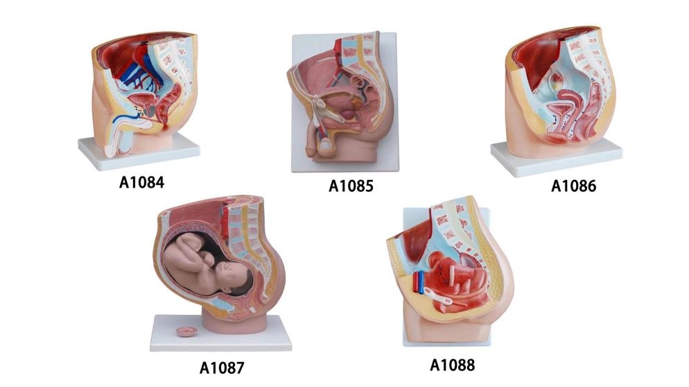
Pelvic Model with Removable Fetus for Midwifery Training
Week Sagittal Section & Removable Fetus
The model gives a detailed look at a female pelvis right at the 40th week of pregnancy, split right down the middle (sagittal section). The best part? The full-term fetus can be completely removed from the uterus for independent study.
Most basic pelvis models are static or sealed shut. Students can see the outside, but they have no idea how tight the fit really is inside.
Professors can take the baby out and pass it around, or manually guide it through the pelvic corridor. It lets students physically feel and see how a full-term baby maneuvers through those tight internal spaces, turning a confusing abstract concept into an instant “aha!” moment.
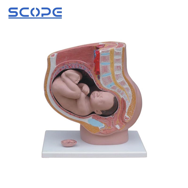
Pre-Birth Fetal Positioning & Complete Urogenital Mapping
This model doesn’t just throw a baby inside a pelvis; it shows the exact, correct fetal position right before birth, sitting alongside the entire female urogenital system.
In a real delivery room, the descending baby puts massive physical pressure on the mother’s bladder and urethra. If a student doesn’t understand this spatial relationship, they risk mismanaging issues like urinary retention or misplacing a catheter during labor.
Because the bladder, uterus, and vagina are all clearly laid out in relation to the fetus, students can see exactly how the anatomy shifts under pressure. It’s the perfect, risk-free tool for teaching proper catheterization and protecting maternal tissues during labor.
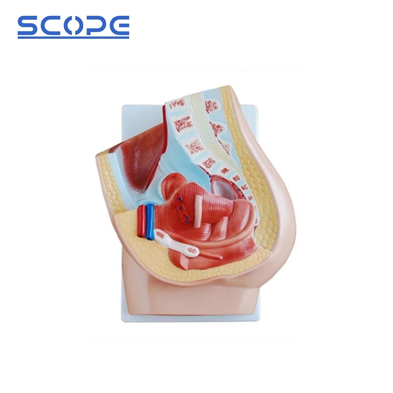
High-Definition Abdominal and Pelvic Floor Muscle Detailing
The sagittal slice provides a crisp, highly detailed look at the abdominal wall and the pelvic floor muscles.
The pelvic floor is notoriously hard to teach because it’s tucked deep inside the bony pelvis. Many students graduate without really understanding how these muscles stretch, flex, or tear when the baby’s head crowns.
Instructors can point directly to the crisp muscle layers to explain the mechanics of maternal pushing forces. It makes it incredibly easy to show students where tears are likely to happen and visually map out the exact path for an episiotomy.
Comprehensive Internal Reproductive Anatomy
Look inside the sagittal section, and you’ll see a clear layout of the female genitalia, uterus, vagina, and the broad ligament of the uterus (ligamentum latum uteri).
A lot of basic simulators treat the uterus like a floating organ, completely ignoring structural supports like the broad ligament. This leaves students blind to how the entire reproductive tract is anchored and how it changes during contractions.
This deep structural view makes the model useful far beyond just standard childbirth classes. It’s an excellent asset for general gynecology and nursing programs to trace the anatomy from the vagina all the way up to the supporting ligaments.
Multi-Stage Curriculum: Integrated 3-Month Embryo Model
To give you more bang for your buck, the sturdy base of this model includes a separate, detailed 3-month-old embryo model for comparative study.
Usually, schools have to buy one expensive kit for early embryology and a completely different one for late-stage labor training. It blows the budget and clutters up the lab counters.
It’s a massive win for procurement and lab space. A professor can use the exact same model to teach early fetal development at the start of the semester, and then pivot to a full-term 40-week delivery later on.
Premium, Durable PVC Construction
The entire model is constructed from high-grade, premium PVC material.
Simulation labs are busy, high-traffic environments. Cheap models chip, warp, or stain after a semester of being handled by hundreds of students, forcing you to constantly re-order.
This heavy-duty PVC build is impact-resistant, non-toxic, and 100% washable. Students can handle it, trace the structures with their fingers, and you can easily wipe it down with disinfectant after class without worrying about fading or cracking. It’s a long-term investment that actually lasts.
How to Choose the Right Model?
If you are a medical distributor or a procurement manager picking out models for a university lab, keep these three criteria in mind to get the best value:
- Life-Sized Accuracy Matters: Always ensure the model is a 1:1 life-sized, anatomically accurate representation. When you are teaching pelvic dimensions and fetal descent, proper scale is absolutely critical—scaled-down models will confuse students.
- Check the Moveable Parts: Make sure the fetus is fully removable and easy to reposition within the pelvic cavity so instructors aren’t struggling mid-lecture.
- Material and Kit Content: Prioritize high-grade PVC for durability. Also, assess whether the department just needs a solid anatomical slice or if they require an expanded kit with a placenta and umbilical cord for a comprehensive birth simulation.

