

At the top of the human spinal column, the first cervical vertebra, C1 (Atlas), and the second cervical vertebra, C2 (Axis), form the most structurally unique and precisely engineered skeletal combination in the body.
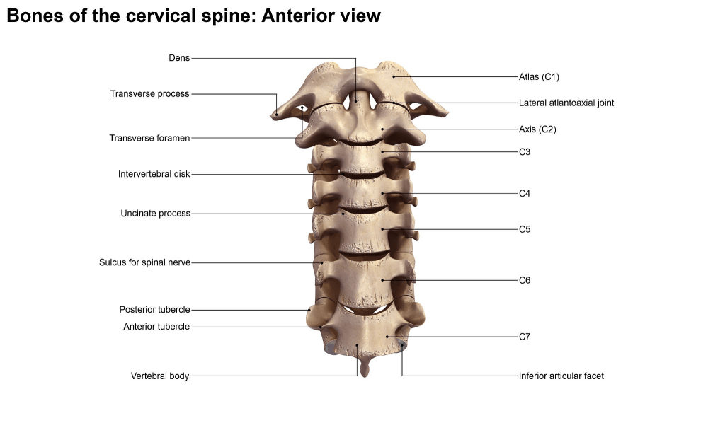
Think of C1 and C2 as a specialized pivot-and-collar assembly at the top of your neck. Working together, they allow your head to perform complex movements while supporting its considerable weight:
- C1 (Atlas): Supports the full weight of the skull. It has no vertebral body and no spinous process, and its primary function is facilitating the nodding motion (saying “Yes”).
- C2 (Axis): Serves as the central pivot for C1’s rotation. Positioned directly underneath C1, its primary function is facilitating the side-to-side rotation (saying “No”).
| Comparison Dimension | C1 Vertebra (Atlas) | C2 Vertebra (Axis) | Mechanical & Functional Logic |
| Morphology | Bony ring (no vertebral body, no spinous process) | Vertebral body featuring an upward vertical peg (Dens / Odontoid Process) | C1 acts as a “collar” enclosing C2’s “pivot pin”. |
| Primary Function | Directly cradles the base of the skull | Provides the axis for horizontal head rotation | Combined, they ensure multi-directional head mobility. |
| Dominant Movement | Pitching / Nodding (“Yes” movement) | Swiveling / Shaking (“No” movement) | The C1-C2 joint contributes roughly 50% of total head rotation. |
| Joint Formed | Atlanto-occipital joint | Atlanto-axial joint | Lacks thick intervertebral discs; relies on ligamentous and bony alignment. |
C2 Vertebra Model
Primary Applications
- Rotation & Pivot Mechanism Teaching: Demonstrates the C2 Dens acting as an axis that carries C1 and the skull through wide rotation angles (shaking “No” or turning to look sideways).
- High-Risk Fractures & Critical Pathology Explanation: Used to explain trauma caused by car accidents or severe falls, such as Odontoid Fractures and Hangman’s Fractures, as well as the risk of paralysis from high cervical spinal cord compression.
- Advanced Orthopedic Surgical Training: Enables orthopedic surgeons to practice high-difficulty procedures on realistic models, including C2 Pedicle Screw Placement and Cannulated Odontoid Screw Fixation.
Advantages
- Distinct Anatomical Landmarks: The prominent, upward-projecting Dens and large bifid spinous process make it highly recognizable and unmistakable.
- High Mechanical & Educational Value: As the core axis of the upper cervical spine, it is an indispensable component for demonstrating atlanto-axial joint kinematics.
- Essential for Surgical Tactile Training: The narrow pedicles adjacent to the spinal canal make it the ideal physical tool for practicing precise surgical drilling and screw placement.
Disadvantages
- Complex Standalone Geometry: The slender, protruding Dens is more vulnerable to damage or breakage during mold extraction, handling, and transit compared to standard vertebrae.
- High Manufacturing Precision Required: Strict dimensional tolerances are crucial; improper manufacturing can lead to an overly tight fit or loose, unstable movement when assembled with C1.
Limitations
- Cannot Demonstrate Nodding Alone: The superior articular facets of C2 do not interface directly with the skull (occiput), making it impossible to demonstrate forward/backward pitch (“Yes” movement) using C2 alone.
- Cannot Demonstrate Full Rotation Independently: Head rotation requires C1. Without C1, a standalone C2 cannot visually convey the physical mechanism of turning the head.
C1 Vertebra Model
Primary Applications
- Skull Support & Nodding Movement Demonstration: Shows how the skull (occiput) rests upon C1 and demonstrates the forward/backward tilting mechanism (“Yes” movement).
- Specialized Anatomical Structure Teaching: Serves as a classic example of atypical bone morphology—being the only vertebra in the spine without a body or spinous process.
- Pathology & Patient Education (Dizziness / Migraines): Combined with the vertebral artery and greater occipital nerve, it helps explain how C1 misalignment compresses neurovascular structures, leading to cervicogenic dizziness and occipital headaches.
- Fundamental Orthopedic Training: Allows surgeons to practice anatomical landmarking and trajectory angles for C1 Lateral Mass Screw Placement.
Advantages
- Accurate Representation of Ring Anatomy: Clearly displays unique structures (anterior arch, posterior arch, and bilateral lateral masses), making it easily distinguishable from standard vertebrae.
- Excellent Load-Bearing & Interface Demonstrator: The concave superior articular facets cradle the skull model base, serving as a transitional link between the head and spine.
- Durable Structural Integrity: The closed/semi-closed ring shape distributes stress evenly, making it more impact-resistant than the C2 model with its protruding Dens.
Disadvantages
- Lacks Standard Vertebral Landmarks: Without a conventional vertebral body or spinous process, beginners may find it difficult to identify superior/inferior or anterior/posterior orientation without C2 or a skull base.
- High Ligament Dependence: In vivo stability relies heavily on the transverse ligament of the atlas; without a simulated ligament component on the model, C1 cannot hold C2’s Dens securely during standalone demonstrations.
C1 and C2 Vertebrae Model: Which is best for you?
| Target Customer Type | Recommended Model / Configuration | Key Purchasing Considerations |
| Medical & Nursing Schools | C1 + C2 Dissectible Joint Set (with clear anatomical labels) | Focus on anatomical accuracy (e.g., foramina, facet curvature) and material durability. |
| Spine Surgery & Orthopedic Hospitals | High-Density Simulated Bone C1/C2 Models (drillable for screw placement) | Focus on tactile feedback (bone density) and strict pedicle/lateral mass tolerances for pre-op planning and screw fixation practice. |
| Physical Therapy, Acupuncture & Rehabilitation Clinics | Dynamic Soft-Tissue Model (Occiput + C1 + C2 + Nerves & Vessels) | Focus on visual clarity (demonstrating neurovascular compression) to improve patient communication regarding dizziness and headache origins. |
| Medical Distributors & Foreign Trade | 1:1 Life-Size Full Spine Model (including functional C1/C2 segment) | Focus on cost-effectiveness, packaging safety, and fracture-proof protection for shipping. |