
News
What Fluorescent Dyes Can a Fluorescence Microscope Detect?
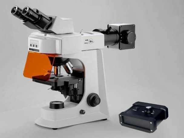
A fluorescence microscope can recognize a wide range of fluorescent dyes, with wavelengths ranging from ultraviolet to infrared light. It’s more about how capable the instrument is of recognizing these dyes, based on its: This means that, as long as the fluorescent dye corresponds with the microscope’s wavelength range, it will work. Classification of Common…
How to Select Filter Cubes Based on Fluorescent Dyes?
Several factors contributing to poor fluorescent signals may include the inappropriate choice of filter cubes for the fluorescence microscope. This rationale could not be simpler: the excitation filter should correspond exactly to the dye excitation wavelength; the emission filter should contain the emission wavelength range, and finally, the dichroic mirror should effectively split the beam….

What are the Essential Features of a High-quality Infant CPR Manikins With Feedback Doll?
When buying training equipment for the CPR training market in 2026, you have to pay attention to whether your training will be in line with the current AHA (American Heart Association) requirements. What are the critical elements of an infant CPR manikin with feedback doll? Simply put, an infant CPR manikin should provide feedback, resist…

Where to Rent Red Cross CPR Manikins Tulsa Oklahoma?
As someone who runs a CPR certification course or a company based in Tulsa, Oklahoma, you know that the first step toward getting ready for such an event involves the search for equipment rentals. Wherever you live in the area, whether close to Broken Arrow, Jenks, or the Tulsa Arts District, manikins from the Red…
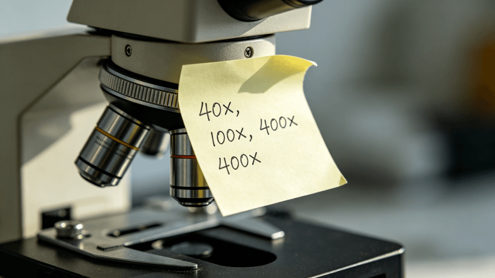
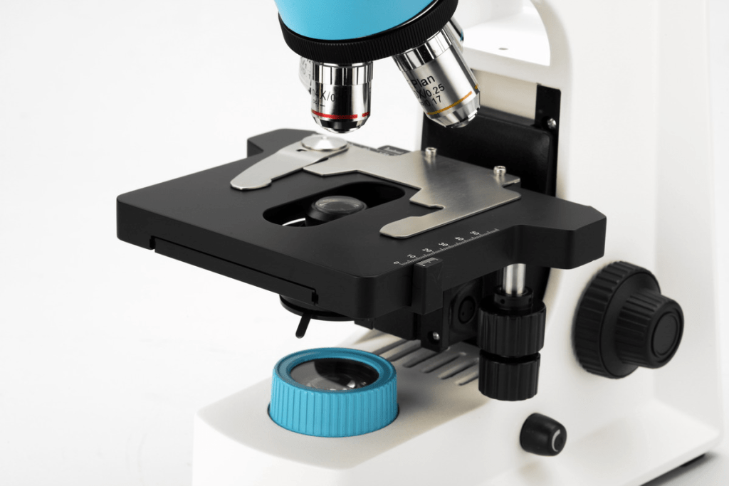
How to Determine the Total Magnification of a Microscope?
There is no need for complex thoughts on magnification or stopping every time to calculate manually. You can achieve the desired accuracy by following this easy trick; just jot it down for easy reference: Here’s one final tip that will help you save even more time: place a strip of tape on your microscope with…
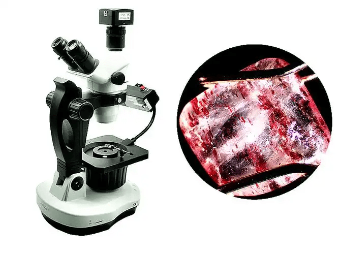
DIY Forensic Lab: How to Detect Synthetic Gemstones and Glass Fakes Using a Polarizing Microscope?
With the sophistication of artificial gemstones these days, one needs more than just a jeweler’s loupe for inspection. Modern synthetics and high lead-glass fakes are made to trick the unaided eye. What is needed is a way of analyzing the behavior of the light itself on a molecular level. For those who have a Polarizing…
Is a $50 USB microscope actually a ‘microscope’ or just a webcam?
Is a $50 USB microscope actually a ‘microscope’ or just a webcam?
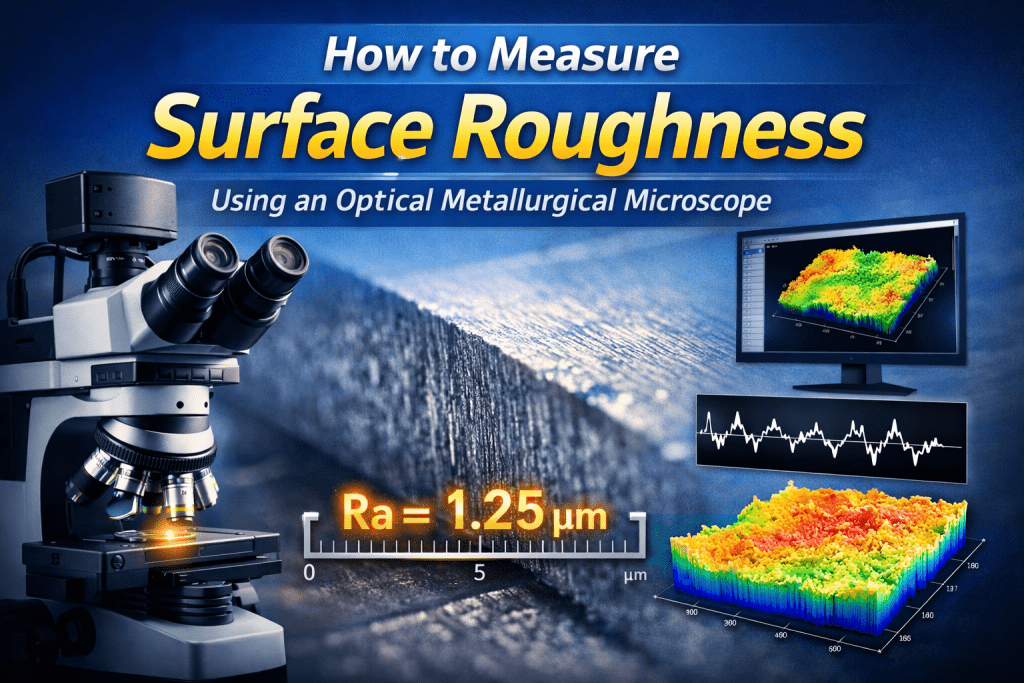
How to Measure Surface Roughness Using an Optical Metallurgical Microscope?
How to Measure Surface Roughness Using an Optical Metallurgical Microscope?
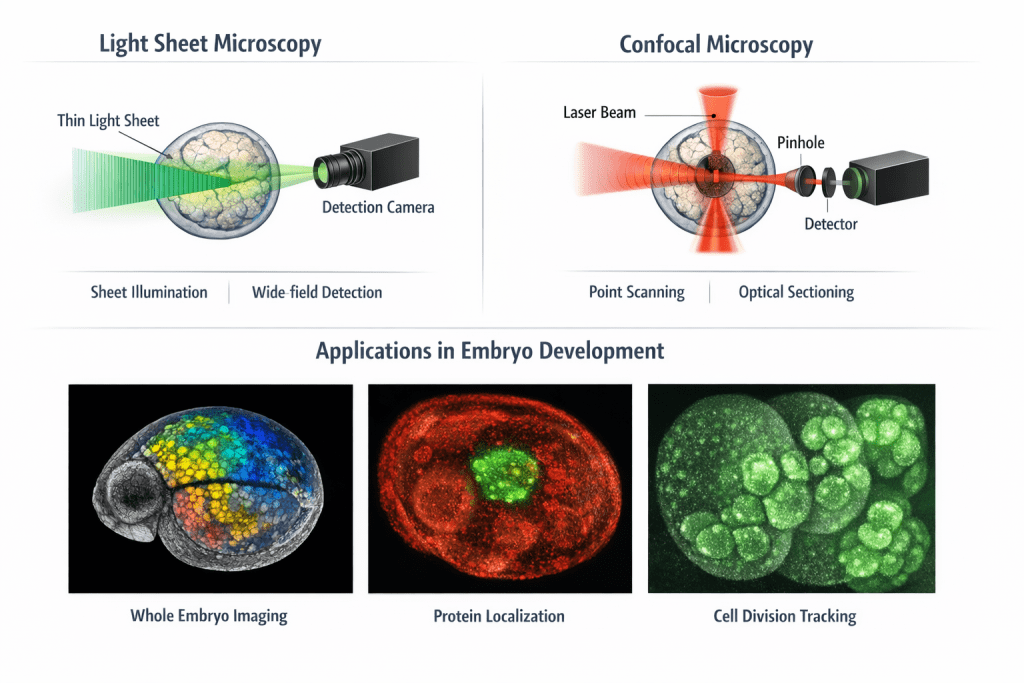
Light Sheet Microscopy vs. Confocal: Which one wins for embryo development tracking?
Light Sheet Microscopy vs. Confocal: Which one wins for embryo development tracking?
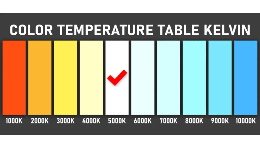
What Color Temperature (K) Is Best for Biological Sample Photography?
In biological photography, getting the color temperature right isn’t just about making things look “pretty.” It’s about data integrity. If your white balance is off, your staining intensity looks wrong, and your peer-reviewed paper suddenly looks a lot less credible. This is the industry standard for “Daylight.” It’s neutral, it’s crisp, and it doesn’t lean…
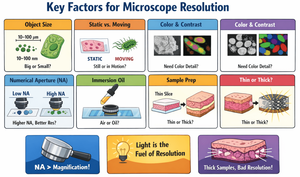
Stop Chasing Magnification: What Resolution Do I Need for Microscopy?
Stop Chasing Magnification: What Resolution Do I Need for Microscopy?
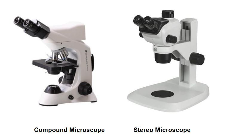
Stereo vs. Compound Microscopes: Which One Should You Actually Buy?
If you’re shopping for a microscope, you’ve probably hit a wall of technical jargon: dual-light paths, numerical aperture, total magnification… But let’s be real—you don’t want a physics lesson. You want to know one thing: “Which of these will help me see what I need to see without wasting my money?” The truth is, these…

