
An anatomically accurate flexible female pelvic model is a key dynamic educational tool designed to simulate the structure, movement, and functional mechanics of the female pelvic region. Unlike traditional rigid plastic models, this flexible model uses realistic, compliant materials that mimic natural joint articulation and soft tissue behavior.
- Pelvic Bones: Ilia, ischium, pubis, sacrum, and coccyx.
- Detachable Pelvic Floor Muscles: High-grade, flexible muscle inserts.
- Reproductive Organs: Uterus, ovaries, and fallopian tubes.
- Viscera: Bladder and rectum (included in advanced anatomical models).
Anatomical Pelvic Model Brands for Patient Education
Scopelab produces a range of medical anatomical models for clinical training and patient education. Their relevant models for pelvic health include:
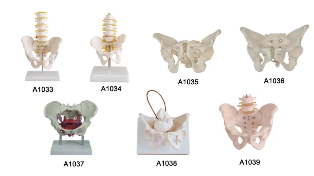
SC-A1037 (Female Pelvis Model with Floor Muscle): Directly features the pelvic floor muscle structures (such as levator ani and urogenital diaphragm) integrated into a natural-sized female pelvic skeleton, making it well-suited for patient education on pelvic floor dysfunction and Kegel exercises.
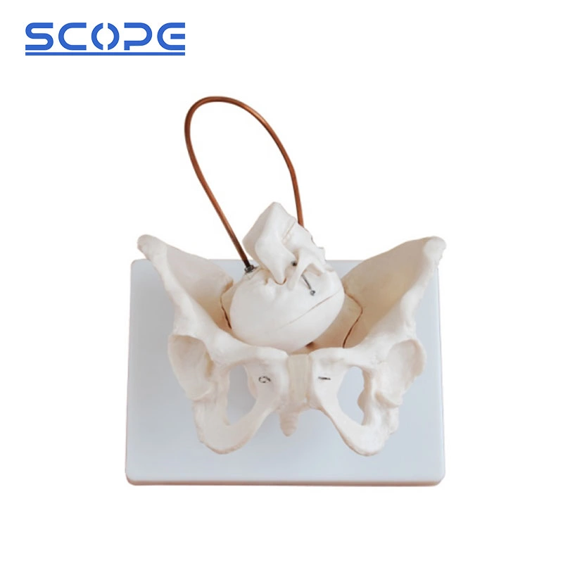
SC-A1038 (Female Pelvis Model with Fetal Skull): Combines the female pelvis with a flexible fetal skull on a jointed rod, useful for demonstrating fetal passage during childbirth alongside pelvic floor mechanics.
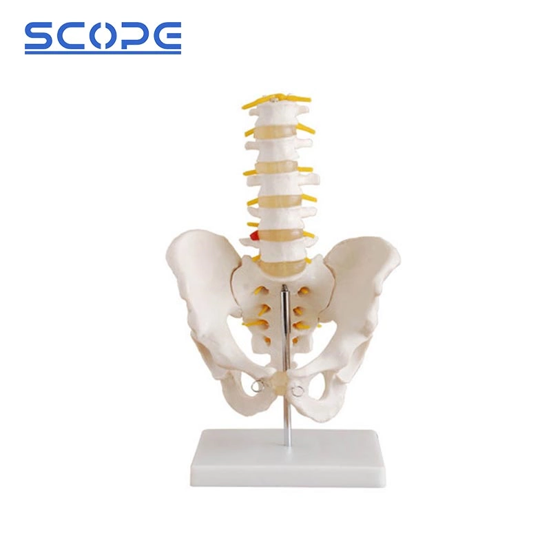
SC-A1033 & SC-A1036: Life-sized basic female pelvis models (with lumbar spine additions available) for focusing on bony landmarks and sacral joints
| Brand | Model Highlight | Ideal Use Case | Key Features |
| Scopelab | SC-A1037 | Educational & Institutional Sourcing | Life-size female pelvis with floor muscles; options for fetal skull demonstration. |
| 3B Scientific | H20 Series | Clinical & University Settings | Modular, highly durable, and features digital 3D app compatibility. |
| Erler-Zimmer | Flexible Pelvis Line | Physical Therapy & Midwifery | Soft, highly flexible muscle inserts with flexible SI joints. |
| Intimate Rose / GPI | Specialized Patient Models | Patient-Facing Consultations | Soft-touch silicone muscles designed specifically for easy patient interaction. |
Why Detachable & Flexible Pelvic Floor Muscles Matter
1. Motion Beyond Static Anatomy
Unlike fixed models, flexible materials allow live demonstration of biomechanical stress:
- Sacroiliac (SI) Joint & Pubic Motion: Demonstrates dynamic shifts during pregnancy, childbirth, and pelvic asymmetry.
- Pelvic Tilting: Visualizes how anterior and posterior pelvic tilts affect posture, core stability, and lower back alignment.
2. Layer-by-Layer Deconstruction
- Isolating Muscle Layers: Snap-on or magnetic fittings let clinicians strip away superficial perineal muscles to reveal deep pelvic floor structures.
- Hypertonicity vs. Hypotonicity: Physical therapists can bend and stretch muscle inserts to contrast tight, spasming muscles against weak, stretched support layers.
Key Advantages Over Traditional Models
Dynamic Demonstration: Simulates real human movement rather than holding a fixed position.
Higher Patient Engagement: Interactive, tactile learning improves information retention.
Layered Exploration: Allows step-by-step breakdown of anatomy without visual clutter.
Versatile Application: Ideal for both high-level professional training and everyday clinical education.
Who Should Use This Model?
Obstetricians and Gynecologists (OB/GYNs)
Pelvic Floor Physical Therapists & Rehabilitation Specialists
Urologists & Urogynecologists
Medical Educators & Nursing Schools
Sexologists & Health Counselors
Buyers’ Guide & Key Considerations
Medical-Grade Materials: Ensure bones are cast from high-durability PVC and muscle layers are made of washable, eco-friendly silicone built for frequent handling
Magnetic vs. Snap Attachments: Magnetic fittings allow seamless, quick disassembly during brief 15- to 30-minute consultations.
Integrated Spine & Femur Options: Models that include L1–L5 lumbar vertebrae or removable femoral heads offer a complete view of the lower extremity kinetic chain.