

Choosing the right teaching tool is essential for effectively demonstrating human anatomy and biomechanics. When selecting between a mini spine model (typically half-scale, around 15–18 inches) and a full-size spine model (1:1 life-size scale, around 32–35 inches, often including the pelvis and femur heads), the decision largely comes down to your teaching environment, audience size, and mobility requirements.
Both models serve distinct purposes—ranging from university anatomy laboratories and chiropractic clinics to mobile physical therapy sessions:
- Mini Spine Models (1/2 Scale): Best suited for small-group demonstrations, patient education handouts, mobile practitioners, one-on-one desk consultations, or programs operating on a tight budget.
- Full-Size Spine Models (1:1 Scale): Best suited for formal university anatomy courses, large-group lectures, and advanced demonstrations of biomechanical dysfunctions and spinal pathologies.
| Feature | Mini Spine Model (1/2 Scale) | Full-Size Spine Model (1:1 Scale) |
| Height | ~15–18 inches (38–45 cm) | ~32–35 inches (80–90 cm) |
| Portability | Highly portable; fits easily into a backpack or small bag | Bulky; usually remains on a dedicated floor or desk stand |
| Anatomical Realism | Good overall curvature and major structures; scaled down | Exact 1:1 scale; accurate vertebral bodies, spinal nerves & discs |
| Visibility in Groups | Best for 1-on-1 or small-group desk demonstrations | Clearly visible across a full classroom or lecture hall |
| Biomechanics & Flexibility | Demonstrates basic curves and bending; fine movement can be stiff | High fidelity for demonstrating disc herniations, subluxations & range of motion |
Mini Spine Model Best For: Mobile educators, desk consultations, student home study, or compact clinical spaces.
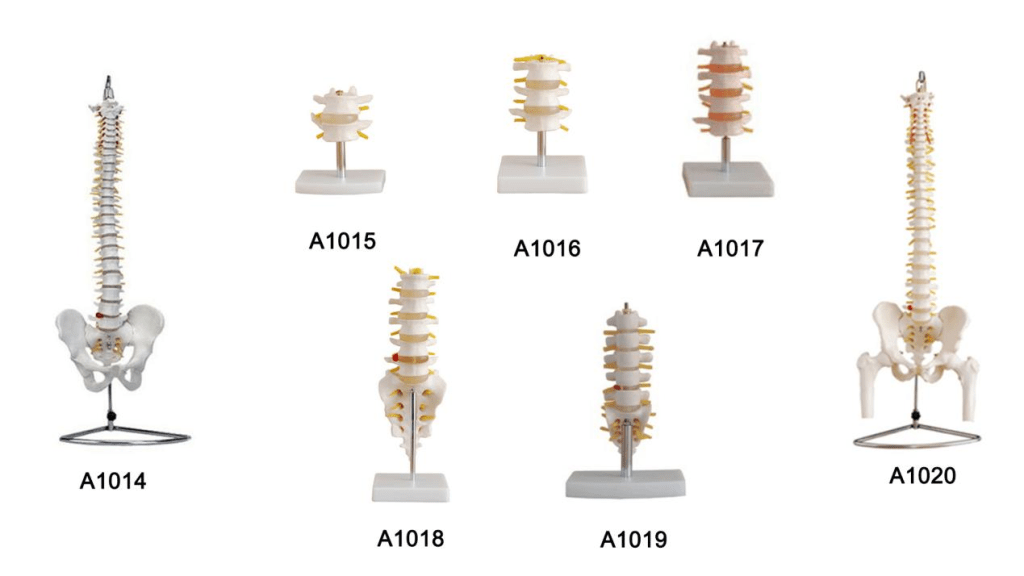
Pros:
- Ultra-Portable: Easily carried between treatment rooms, satellite clinics, or home visits.
- Less Intimidating: Takes up minimal desk space during patient consultations.
- Budget-Friendly: A cost-effective solution when outfitting multiple student lab stations.
Cons:
Limited Detail: Smaller features (such as facet joints, nerve roots, and spinal cord branches) are hard to see from a distance.
Tactile Limitations: The smaller scale makes demonstrating complex manual therapy techniques or specific vertebral rotations less realistic.
When to Choose a Mini Spine Model?
- Space-Constrained Environments: Outpatient consultation rooms, tutoring kiosks, or mobile teaching kits where a full-size stand is impractical.
- Small-Group or 1-on-1 Education: Chiropractic/physical therapy consults, nursing skills stations, or student study cubicles.
- Budget-Sensitive Programs: Equipping multiple lab stations is far more financially feasible with miniature models.
- Focused Regional Study: Highly effective if your lessons focus on specific cervical, thoracic, or lumbar segments rather than the full axial skeleton as a whole.
High-quality mini models still accurately display vertebrae, intervertebral discs, the sacrum, and often a herniated disc segment, making them exceptionally practical for foundational anatomy and patient education.
Full-Size Spine Model Best For: University biology and anatomy classrooms, physical therapy schools, chiropractic colleges, and lecture halls.
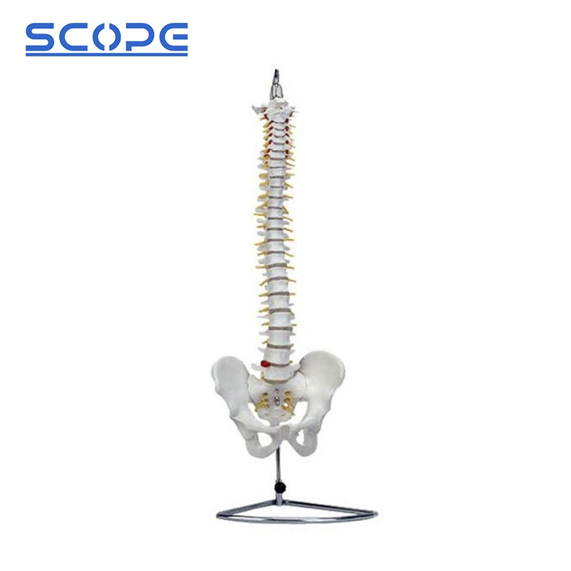
Pros:
- Life-Size Realism: Students can interact with vertebrae at actual scale, making landmark palpation and identification far more realistic.
- Dynamic Demonstrations: Excellent for demonstrating nerve impingement, prolapsed discs, spinal curvatures (scoliosis, lordosis, kyphosis), and pelvic movement.
- Group Visibility: Clear enough for demonstrations in front of a full room or lecture hall.
Cons:
Storage & Weight: Heavy and cumbersome to transport without a dedicated carrying case.
Higher Cost: Premium flexible full-scale models featuring muscular or neurovascular overlays require a larger initial investment.
When to Choose a Full-Size Spine Model:
- Lecture Hall Visibility: Measuring around 85 cm (including the pelvis and femur heads), anatomical landmarks and nerve paths remain visible even from the back of the room.
- Detailed Anatomical Instruction: Life-size models typically feature comprehensive structures such as the spinal cord, nerve roots, vertebral arteries, vertebral notches, and pathological landmarks.
- Hands-On Clinical Skills Training: For practicing pedicle screw instrumentation, manual therapy techniques, or biomechanical adjustments, the 1:1 scale provides authentic tactile feedback and alignment verification.
- Comprehensive Teaching: Essential when you need to demonstrate full-spine curvature, lumbopelvic alignment, and segmental relationships simultaneously.
- Recent advancements in 3D-printed life-size spine models have also demonstrated that high-fidelity replicas can maintain accurate morphology and functional mobility for hands-on medical training.