
Stereo microscopes display larger 3D objects at low magnification, allowing for observation and hands-on operation, whereas compound microscopes display very small, thin samples at high magnification, allowing for the observation of cells and fine internal structures.
| Features | Stereo Microscope | Compound Microscope |
| Also called | Dissecting microscope | Biological / High-power microscope |
| Typical magnification | Low (typically 10x to 40x) | High (typically 40x to 1000x or higher) |
| Image type | True 3D upright view | Two-dimensional (2D) flat image |
| Optical path | Two independent optical paths (one for each eye) | Single shared optical path split into two eyepieces |
| Lighting | Reflected light (illuminates the surface of the object) | Transmitted light (passes through the sample from below) |
| Specimens | Large, opaque objects (e.g., insects, circuit boards, coins) | Thin, transparent slides (e.g., cells, bacteria, thin tissue sections) |
| Working distance | Long, leaving space for placing tools | Short, especially at high magnification |
| Best suited for | Dissection, electronics, entomology, mineralogy, soldering | Cells, bacteria, tissue sections, microstructures, life sciences |
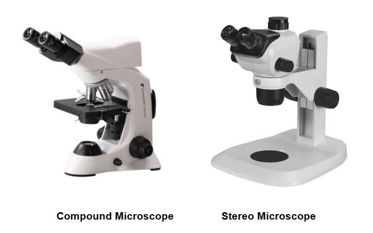
Magnification and Image Depth (2D vs. 3D)
Stereo microscope: Focuses on lower magnification (10x to 40x). It does not pursue extreme detail, but instead provides a wide field of view and true depth perception. It uses two independent optical paths (one for each eye), transmitting slightly different-angle images to the brain, thereby forming a true 3D image.
Compound microscope: Focuses on high magnification (40x to 1000x or higher). It utilizes a series of objective and ocular lenses to magnify the observed sample. Even with two eyepieces, the image seen by both eyepieces is completely identical, so the final presentation is a two-dimensional flat image.
Illumination and Specimen Preparation (Reflected vs. Transmitted)
The way light hits the object changes what you see:
Reflected light (Stereo): Light shines directly onto the top or sides of the sample. This makes it ideal for opaque solid objects that light cannot penetrate, such as coins, circuit boards, insects, or plant leaves.
Transmitted light (Compound): Light shines from underneath and passes directly through the specimen. Therefore, the specimen must be very thin and transparent (e.g., blood smears, cheek cells, or thin plant sections placed on a glass slide).
Working Distance and Hands-on Operation
Stereo microscopes have a large working distance—that is, the physical distance between the bottom objective lens and the stage. This provides ample space for soldering micro-components, dissecting specimens, or manipulating samples with tweezers and scalpels.
Compound microscopes have a very short working distance, especially at 40x or 100x oil immersion objectives, where the distance between the lens and the glass slide is only a fraction of a millimeter.
Stereo Microscope vs. Compound Microscope: Which One Should You Choose?
Stereo Microscope: Ideal for macroscopic objects that require physical manipulation, dissection, soldering, or detailed surface inspection.
Compound Microscope: Ideal for cellular and microbiological studies where you need to examine internal cellular structures, microorganisms, or thin slide-mounted specimens.
Compound vs Stereo Microscope: Which One You Need?
How Does a Compound Microscope Differ from a Stereo Microscope?