Description
I. Introduction to the Upper Respiratory Tract Simulation Model
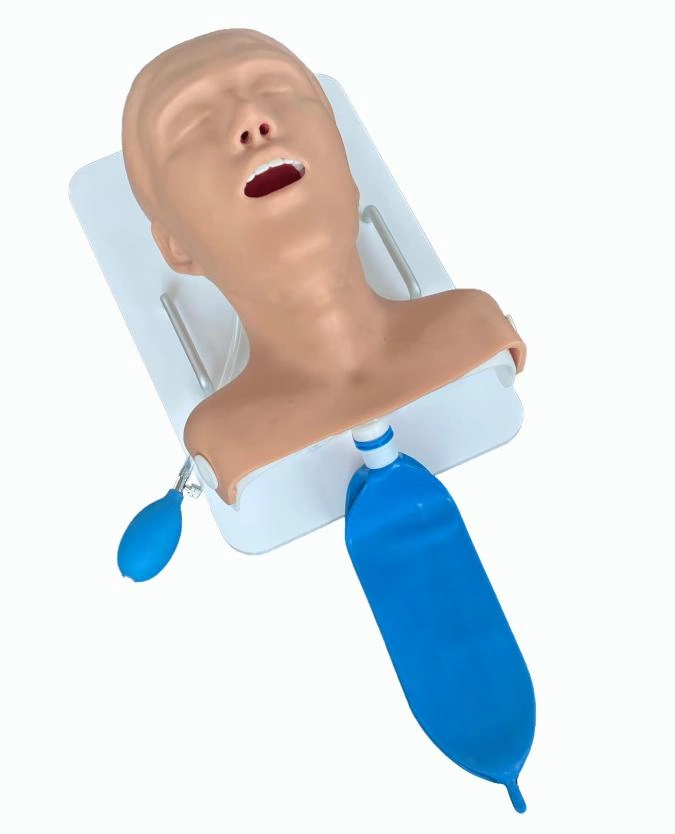
The upper respiratory tract simulation model is a complete respiratory tract training model. It allows medical students to understand the anatomical morphology and structure of the respiratory tract, learn airway management operations, experience the actual operation of the laryngeal mask and laryngoscope, and obtain realistic visual and tactile sensations during surgery, enabling doctors to learn better and faster.
II. Surgical Training Procedures
1. Learning about Respiratory Tract Structure
2. Laryngeal Mask Operation Training
3. Laryngoscopy Operation Training
4. Airway Management Operation Training
III. Upper Respiratory Tract Simulation Model
1. Specifications and Dimensions: 30*23.3*48.8CM
2. Adult physique appearance, based on real human image data, 1:1 realistic and complete human anatomical structure, 1:1 human organ cavity curvature and path, supine human model, maintaining head in a neutral position.
It includes the physiological structures of the skin, skull, trachea, esophagus, nose (nasal vestibule, superior, middle and inferior turbinates, superior, middle and inferior nasal meatuses, nasal septum, eustachian tube torus, eustachian tube orifice), mouth (lips, teeth, tongue, tongue root, hard palate, soft palate, uvula, palatopharyngeal arch, palatine tonsils), and larynx (epiglottis, laryngeal orifice, vocal folds, pyriform fossa, esophageal orifice).
3. The internal airway structure is based on a real human CT scan model, including the complete nasal cavity, nasal septum, inferior turbinates, middle turbinates, uvula, tongue, epiglottis, pyriform fossa, slit, and glottis. A separate opening to the esophagus is connected to the pyriform fossa, which can be used to place a gastric tube and a double-tube laryngeal mask airway.
4. The exterior features important head features such as the mouth, nose, eyes, eyebrows, ears, and hair. Openings are located at corresponding positions in the mouth and nose for airway management.
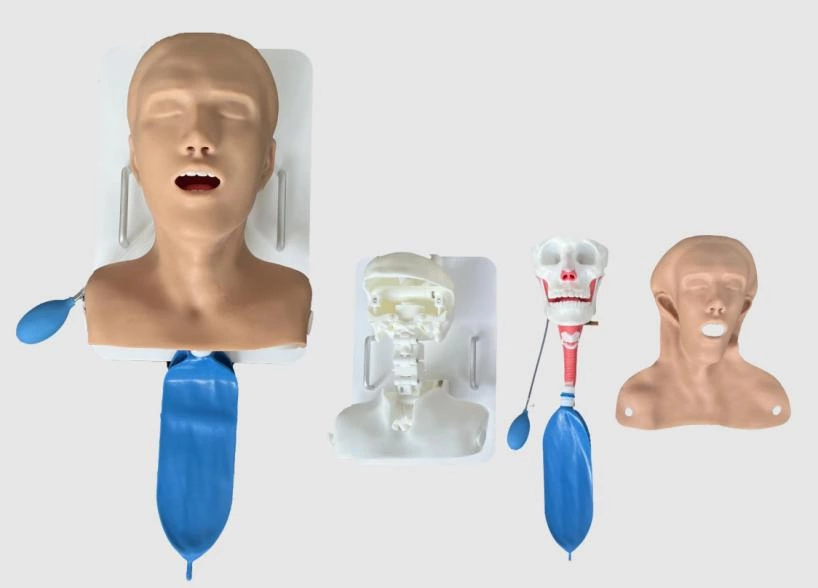
5. The mandible is independently 3D printed, with the condyle of the mandible connected to the mandibular fossa of the temporal bone by a highly elastic elastic band. This accurately reproduces normal movements such as opening and closing the mouth, mandibular forward movement, and rebound, allowing for mandibular lifting actions.
6. The outer skin is fixed to the model’s bony structure by locking points on the eye sockets and both shoulders, allowing for easy removal and replacement.
7. Some cervical vertebrae are independently 3D printed, enabling cervical joint movement. By adjusting the position of the posterior occipital bone limiter, the model can achieve neutral, tilted, and hyperextended neck positions. The mouth opening of the model also changes in tandem with these positional changes.
8. The trachea is connected posterior to the glottis. The membranous part of the trachea and the tracheal cartilage rings are realistically reproduced. The anterior surface of the trachea features bony landmarks of the thyroid cartilage and cricoid cartilage. By applying pressure to the cricoid cartilage, the airway position can be changed, the esophagus can be closed, and cricoid cartilage compression (Sellick technique) can be practiced. *9. The airway structure is detachable and replaceable.
10. The tongue contains an inflatable balloon, which can be easily inflated to recreate difficult airway conditions such as an enlarged tongue or tongue swelling. It can be deflated with a single button.
11. The model surface uses a dust-proof process, making it easy to wipe and preventing dust from adhering.
12. The outer skin is the skin tone of a typical Chinese person, with a Shore hardness of 0HA. It has a certain thickness and toughness, effectively replicating the feel and elasticity of real human skin and tissue.
13. The internal airway material simulates the color of real human tissue, being red, with a Shore hardness of 10HA. It effectively replicates the feel and elasticity of real human tissue.
14. The skin and airway are made of a specially formulated platinum silicone biomimetic material, which is safe, non-toxic, environmentally friendly, and odorless. It can be marked for positioning and wiped multiple times; it can be used for puncture, cutting, suturing, and cannulation. Subcutaneous surgical procedures feel realistic.
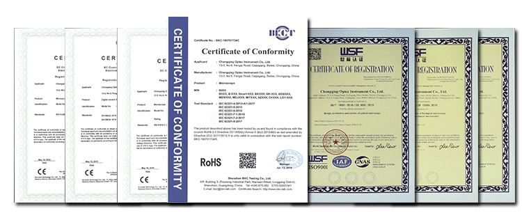
15. RoHS official non-toxic testing certificate is available.
16. The bony parts of the model are modeled entirely according to the normal human head and face skeletal structure and 3D printed. The nasal cartilage is 3D printed separately and used for shaping the bridge of the nose.
17. It can be used to understand the anatomy and structure of the upper respiratory tract and to learn and experience the actual operation of laryngeal masks and laryngoscopes.
Supports both nasal and oral insertion methods. Practice of both endotracheal intubation methods is possible. Lubricant is required; apply medical lubricant to the tool for easy airway entry.
18. Supports external laryngeal manipulation, cricoid cartilage compression (Sellick technique), cricothyroid membrane localization, puncture, and other operations.
19. Both flexible and rigid endoscopes can be used for training.
IV. Basic Configuration Requirements
1. Upper respiratory tract simulation model *1
2. Fixing base *1
3. Fixing bracket *1
| Why Choose Us?
SC-LV40 medical training ear washing manikin for ear syringing has relatively specific functions and is a good simulator for training medical staff’s ear canal irrigation skills. The service life of the product is relatively long, and it can be trained many times without deformation. We all provide a one-year after-sales guarantee service, and technicians will solve after-sales problems for customers online. Our price is moderate, and we will customize logos for customers according to their needs. There will be no additional charge for domestic shipping.
| Order Process
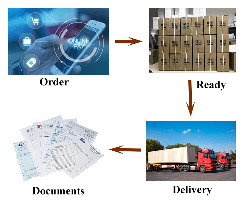
- After confirming the product quantity and shipping method, pay according to the contract.
- After we receive the payment, we will place an order with the factory for production.
- The product is produced and packaged, and shipped by the agreed mode of transportation.
- Confirm the customs clearance documents and product delivery pictures with the customer.
| Company
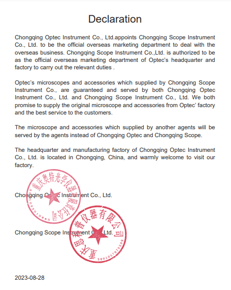
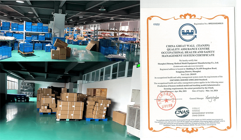
Chongqing Scope was established in Chongqing in 2019. The main business scope is the export of laboratory products. Our sales staff are all experienced employees, we have our own dedicated microscope factory, and products can be customized according to requirements. We also provide other laboratory consumables, metallographic equipment, medical simulators, etc. Our customers are located in more than 30 countries and regions all over the world, and we get a lot of good feedback. Our company has various certificate qualifications, ISO9001, ISO14001, CE, UKCA, ISO45001, ISO13485, etc.